
|
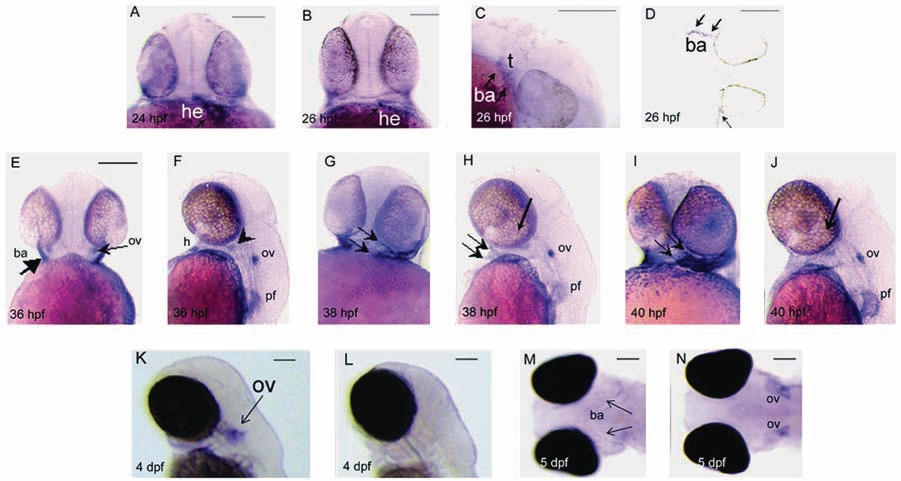
Fig. 4 WISH analysis of wnt9b in developing embryos. (A) Frontal view, 24 hpf embryo. Expression of wnt9b in the early heart tube (arrow, he). (B) Frontal view, 26 hpf embryo. wnt9b expression is restricted to the anterior portion of the heart tube (arrow, he). (C) Lateral view at 26 hpf, anterior to the right. wnt9b is expressed in first and second arch tissues (arrows, ba) surrounding the trigeminal (t) ganglion. (D) Oblique longitudinal sectioned 26 hpf embryo reveals wnt9b expression in branchial arch ectoderm (arrows, ba). (E, F) 36 hpf embryo. Frontal view reveals wnt9b expression in anterior branchial arch tissues (E, arrow, ba), and in the otic vesicles (ov). (F) Lateral view reveals wnt9b expression in the heart (h), otic vesicles (ov), pectoral fin bud (pf), and anterior arch tissue (arrowhead). (G) Frontal view of 38 hpf embryo reveals distinct wnt9b expression in anterior ba tissues (arrows), and lateral view (H) reveals wnt9b expression in ba (arrows), ov, and pectoral fin (pf). (I, J) Branchial arch, ov, and pf wnt9b expression persists at 40 hpf. At 4 dpf (K, arrow) and 5 dpf (M, N), wnt9b is detected in otic vesicles, but is reduced or absent in branchial arch tissues. (L) All sense controls were negative. Scale bars, 100 μm.