
|
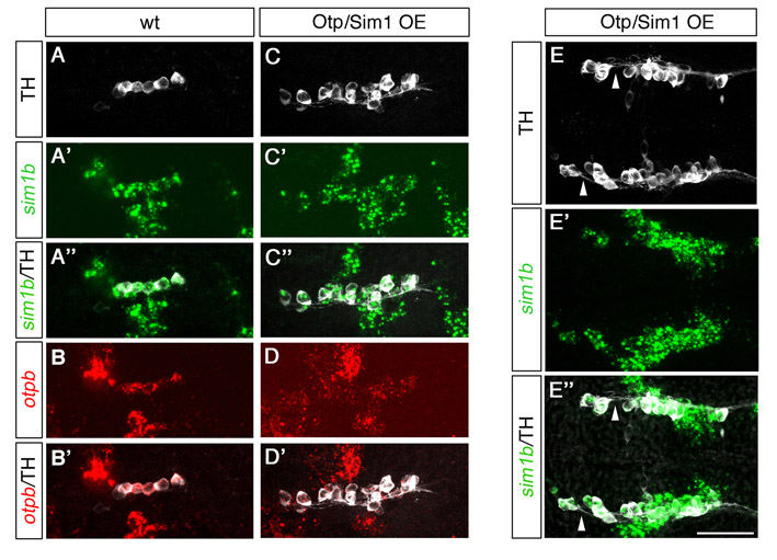
Fig. S11 Supernumerary DA neurons in Otp/Sim1OE embryos express both otp and sim1. (A-E″) Expression of sim1b (whole-mount in situ hybridization, green), otpb (whole-mount in situ hybridization, red) and TH (immunostaining, white) at 25 hpf in wild-type control (A-B′) and transgenic embryos expressing otpa from the hsp70 promoter co-injected with sim1a mRNA (Otp/Sim1OE; C-E″). Lateral view of the region of DA specification in the posterior tuberculum/hypothalamus in wild-type embryos showing co-expression of TH with sim1b (A-A″) as well as with otpb (B-B′). Lateral view of the same brain region in an Otp/Sim1OE embryo showing supernumerary ectopic TH cells (total of 16 cells) that are arranged in a rostrocaudally elongated expression domain but still express sim1b (C-C″) and otpb (D-D′), reflecting the expanded sim1b/otpb expression domains. (E-E″) Dorsal view of TH cells in an Otp/Sim1OE embryo showing that ectopic, anteriorly located TH/sim1b-positive cells send projections caudally where they join the regular DA axonal tracts (arrowheads in E,E″). Anterior is towards the left. Scale bars: 50 μm. Fluorescent images in represent z-projections from single focal planes: (A-D′) 20 μm; (E-E″) 40 μm.