
|
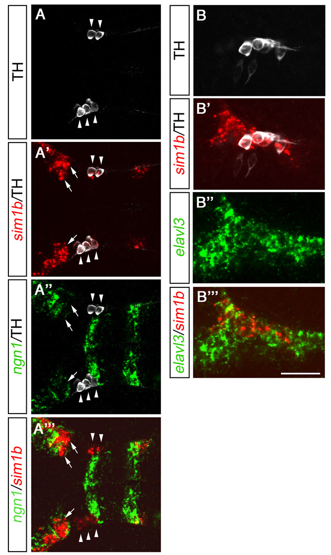
Fig. S7 sim1b is expressed in differentiating neurons. (A-A″′) Detection of neurogenin1 (ngn1; green) and sim1b (red) expression by fluorescent whole-mount in situ hybridization combined with immunohistochemistry for TH (white) at 1 dpf (z-projection, 6 μm). All THir cells co-express sim1b (A′) but not ngn1 (A″; see arrowheads A-A″′). Sim1b expression is only detectable in cells that express very low levels of ngn1 or cell that do not express ngn1 at all (A″′; see arrows A′-A″′). (B-B″′) Fluorescent whole-mount in situ hybridization for elavl3 (huC; green) and sim1b (red) combined with immunohistochemistry for TH (white) at 1 dpf. All sim1b-positive cells co-express elavl3 (B″′). (A-B″′) Lateral views. Anterior is towards the left. Scale bars: 62.5 μm in A-A″′; 50 μm in B-B″″. A-A″′ represent z-projections of single focal planes. Images in B-B″′ represent a single focal plane.