
|
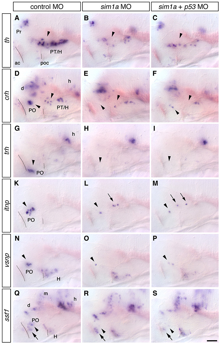
Fig. S6 The loss of specific neuronal subtypes in sim1a morphants is not due to p53-dependent cell death. (A-S) Expression of tyrosine hydroxylase (th, A-C), corticotropin releasing hormone (crh, DF), thyrotropin releasing hormone (trh, G-I), isotocin neurophysin (itnp, K-M), vasotocin neurophysin (vsnp, N-P) and somatostatin1 (sst1, Q-S) at 76 hpf in embryos injected either with 4.5 ng control morpholino (A,D,G,K,N,Q), 1 ng sim1a splice MO alone (B,E,H,L,O,R) or 1 ng sim1a splice MO together with 7 ng p53 ATG MO (C,F,I,M,P,S). The level of reduction in cell number is identical for all analyzed neuronal subtypes in embryos injected with sim1a morpholino alone (arrowheads in B,E,H,L,O,R) or co-injected with p53MO (arrowheads in C,F,I,M,P,S). Arrows indicate ectopic itnp cells (L-M). (A-S) Lateral views; anterior is towards the left. Scale bar: 50 μm. Images represent z-projections from single planes for an better overview. Abbreviations: ac, anterior commissure; d, diencephalon; m, mesencephalon; h, hindbrain; H, hypothalamus; poc, post optic commissure; PO, preoptic region; PT, posterior tuberculum.