
|
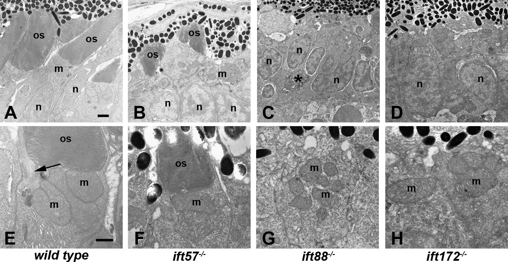
Fig. 3 Transmission electron micrographs of transverse sections along the dorsal–ventral axis of wild type, ift57, ift88, and ift172 mutant zebrafish at 96 hpf. (A–D) Low magnification images revealed longer outer segments (os) in wild type embryos. Photoreceptor outer segments were present in ift57 mutants but were significantly shorter than wild type. Mitochondria (m) and nuclei (n) appear to be normal in the mutants. No outer segments were present in ift88 and ift172 mutants. Nuclei of ift88 and ift172 mutants were less elongated than wild type and pyknotic nuclei (asterisk, C) were occasionally observed. (E–H) At higher magnification, the disk membranes of wild type and ift57 mutant outer segments were highly organized. Arrow indicates connecting cilium. Numerous mitochondria (m) were seen in the apical inner segments of wild type and mutant photoreceptors. Scale bars = 1 μm (A–D), and 500 μm (E–H).
Reprinted from Vision Research, 49(4), Sukumaran, S., and Perkins, B.D., Early defects in photoreceptor outer segment morphogenesis in zebrafish ift57, ift88 and ift172 Intraflagellar Transport mutants, 479-489, Copyright (2009) with permission from Elsevier. Full text @ Vision Res.