
|
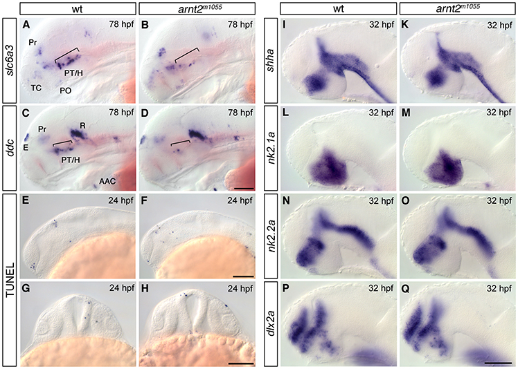
Fig. S2 Forebrain development is largely normal in arnt2m1055 mutants. (A,B) Expression analysis of slc6a3/dat by whole-mount in situ hybridization at 78 hpf revealing reduction of slc6a3/dat-expressing cells in the ventral diencephalon of arnt2m1055 mutants (B) compared with wild-type embryos (A, brackets). Other slc6a3-expressing cells located in the telencephalon, pretectum and preoptic region are not affected. (C,D) Expression analysis of ddc by whole-mount in situ hybridization at 78 hpf revealing reduction of ddc-expressing cells in the ventral diencephalon of arnt2m1055 mutants (D) compared with wild-type embryos (C, brackets) but not in the epiphysis, pretectum, arch-associated neurons or raphe nuclei. (E-H) TUNEL at 24 hpf reveals that apoptosis is not increased in arnt2m1055 mutants (F,H) compared with wild-type siblings (E,G). (I-Q) Expression analysis of shha (I,K), nk2.1a (L,M), nk2.2a (N,O) and dlx2a (P,Q) at 32 hpf reveals no significant change in their expression in arnt2m1055 mutants (K,M,O,Q) compared with wild-type embryos (I,L,N,P). (A-F,I-Q) Lateral views, anterior is towards the left; (G-H) frontal views. Scale bars: in D, 100 μm for A-D; in F, 100 μm for E,F; in H, 100 μm for (G,H); in Q, 50 μm for I-Q. Abbreviations: AAC, arch associated cluster; E, epiphysis; H, hypothalamus; PO, preoptic region; Pr, pretectum; PT, posterior tuberculum; R, raphe nuclei; TC, telencephalic clusters.