
Fig. 4
|
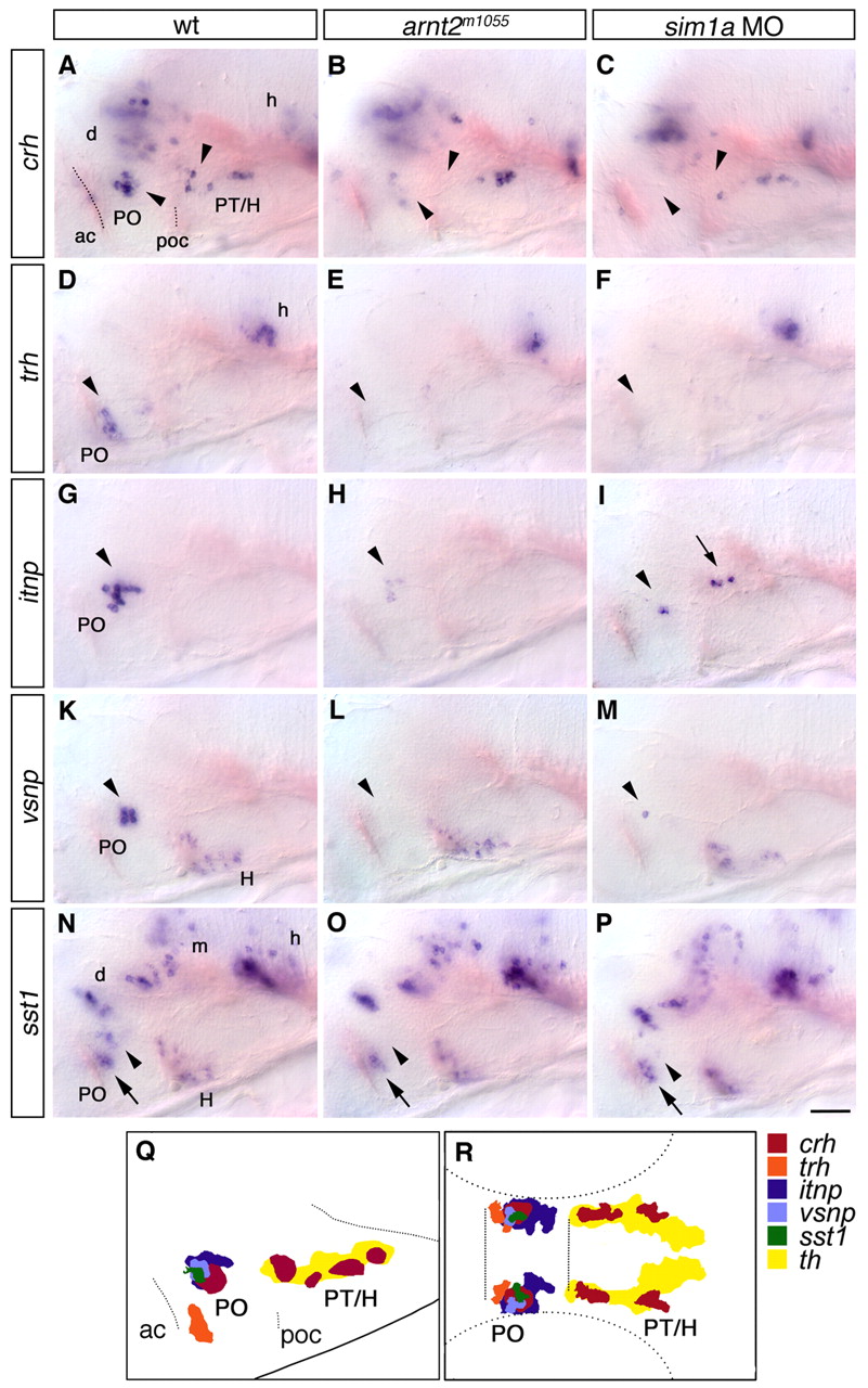
Fig. 4 Reduction of neurohormone-producing cells in the hypothalamus of arnt2m1055 mutants and sim1a morphants. (A-P) Expression of crh (A-C), trh (D-F), itnp (G-I), vsnp (K-M) and sst1 (N-P) at 76 hpf in wild-type embryos (A,D,G,K,N), arnt2m1055 mutants (B,E,H,L,O) and embryos injected with 1 ng sim1a morpholino (C,F,I,M,P). (A-C) crh neurons in the PO (lower arrowheads) and in the posterior tuberculum and hypothalamus (upper arrowheads) are reduced. (D-F) trh-expressing cells in the PO (arrowheads) are strongly reduced. (G-I) itnp-expressing cells in the PO (arrowheads) are strongly reduced or absent. In sim1a morphants, itnp-expressing cells are detected at ectopic locations within the diencephalon (arrow). (K-M) vsnp-expressing cells in the PO (arrowheads) are strongly reduced or absent. (N-P) A group of sst1-expressing cells in the PO (arrowheads) is strongly reduced or absent, whereas all other sst1-positive domains, including a second group in the PO (arrow) are not affected. (Q,R) Camera lucida drawing showing the relative position of all analyzed neuronal groups reduced in arnt2 mutants or sim1a morphants in lateral (Q) or dorsal (R) views; non-affected neuronal groups are not included. (A-P) Whole-mount in situ hybridization, lateral views; anterior is towards the left. Scale bar in P: 50 μm for A-P. Images represent z-projections from several consecutive focal planes. ac, anterior commissure; d, diencephalon; h, hindbrain; H, hypothalamus; m, mesencephalon; PO, preoptic region; poc, post optic commissure; PT, posterior tuberculum.