
|
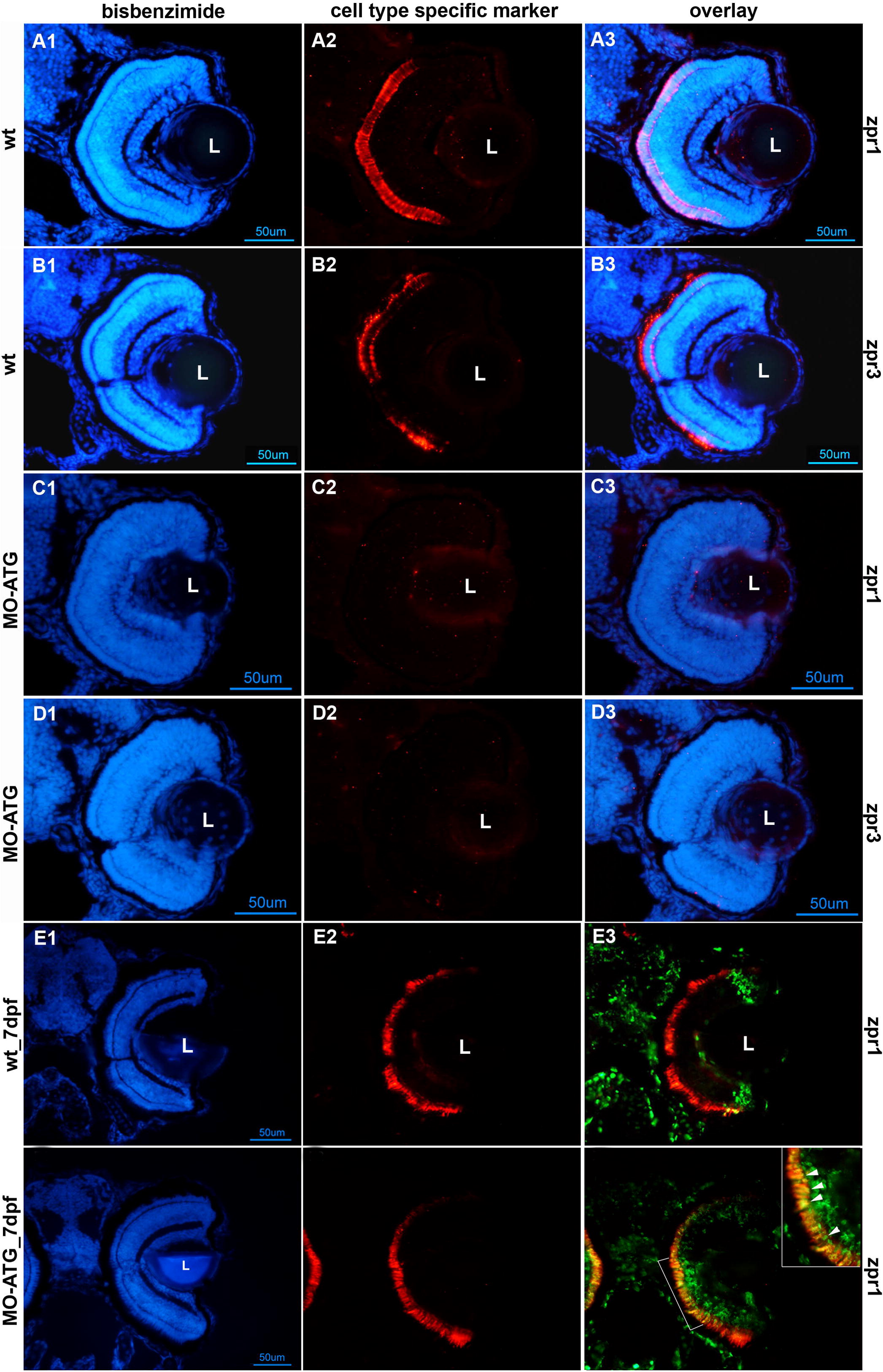
Fig. 8 Photoreceptor differentiation is absent in morphant retinas at 72hpf and recovers in morphant retinas by 7 days post-fertilization. Rows A and B illustrate retinas from wt embryos at 72hpf stained for cone and rod photoreceptors, respectively. Rows C and D are retinas from morphants at 72hpf stained for cone and rod photoreceptors, respectively. Row E illustrates the retina from a wt animal labeled with BrdU at 72hpf, sacrificed at 7days post-fertilization (dpf) and labeled antibodies against BrdU (green) and the cone-specific marker, zpr1 (red). Row F illustrates the retina from a morphant labeled with BrdU at 72hpf, sacrificed at 7dpf and labeled with antibodies against BrdU (green) and the cone-specific marker, zpr1 (red). All left-hand panels illustrate nuclear staining with bisbenzimide. The middle panels are stained with markers for cones (zpr1) or rods (zpr3). The right-hand panels are digital overlays (E3 and F3). The scale bar in panel D1 corresponds to panels A–D and equals 50 μm. The scale bar in panel F1 corresponds to panels E–F and equals 50 μm. L, lens; wt, wild-type embryos; MO-ATG, embryos injected with translation-blocking morpholinos.
Reprinted from Mechanisms of Development, 126(3-4), Ochocinska, M.J., and Hitchcock, P.F., NeuroD regulates proliferation of photoreceptor progenitors in the retina of the zebrafish, 128-141, Copyright (2009) with permission from Elsevier. Full text @ Mech. Dev.