
|
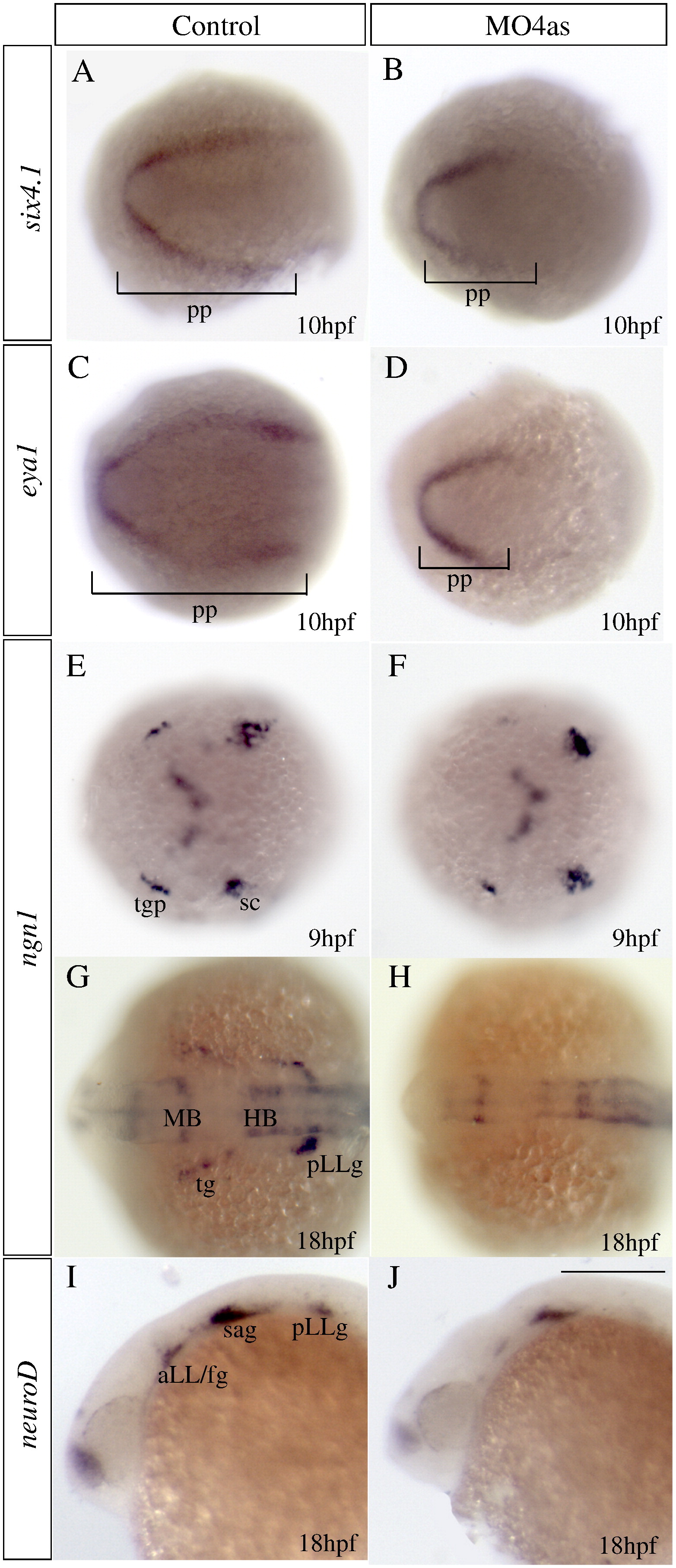
Fig. 4 irx4a is involved in specification of a subset of placodes. In situ hybridization results of embryos processed for detecting expression of the indicated marker genes. Embryos were fixed at 10 hpf (A–F), 17 hpf (G–H) or 24 hpf (I–J). (A–D) Injection of MO4as leads to an inhibition of the posterior expression domain of the preplacodal markers six4.1 (compare A and B) and eya1 (compare C and D). (E–H) ngn1 expression is strongly diminished at 9 hpf in the trigeminal ganglion (tg) but not in the posterior lateral line ganglion (pLLg) in morphants (compare E to F). Later in development, at 17 hpf, expression of this marker is absent in the tg and pLLg (compare G to H). Expression of the proneural gene neuroD in the anterior and posterior lateral line ganglia is also affected in morphants (compare I to J). pp, preplacodal region; aLL, anterior lateral line placode; HB, hindbrain; MB, midbrain; sc, dorsal/lateral spinal cord; sa, statoacoustic ganglion; pLL, posterior lateral line placode; pLLg, posterior lateral line ganglion; tgp, trigeminal ganglion placode; tg, trigeminal ganglion. A–H are dorsal views; I, J show lateral views; anterior is towards the left. Scale bar in J: 40 μm for G–J.
Reprinted from Molecular and cellular neurosciences, 40(3), Feijoo, C.G., Saldias, M.P., De la Paz, J.F., Gómez-Skarmeta, J.L., and Allende, M.L., Formation of posterior cranial placode derivatives requires the Iroquois transcription factor irx4a, 328-337, Copyright (2009) with permission from Elsevier. Full text @ Mol. Cell Neurosci.