
|
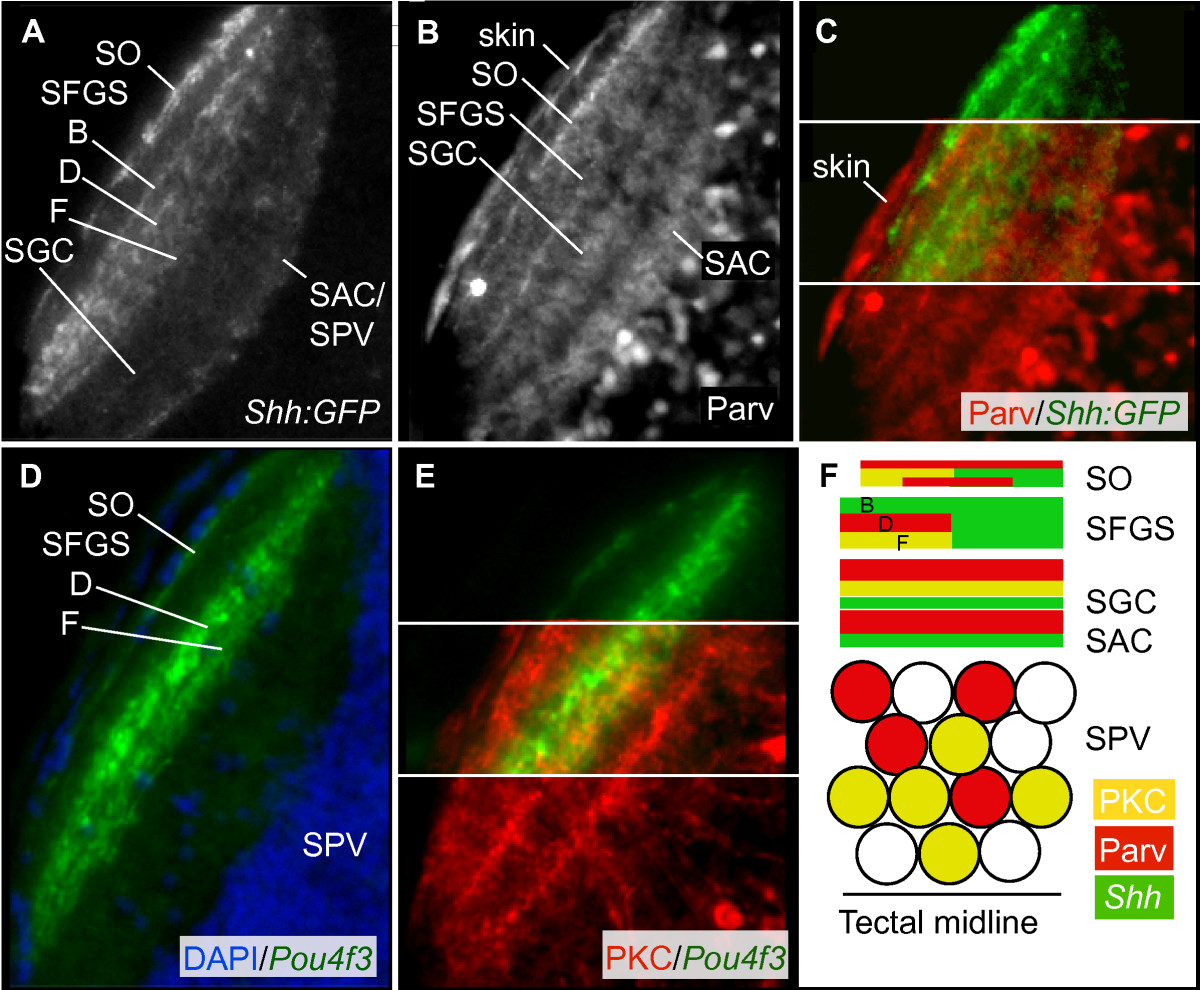
Fig. 2 Neuropil organization of the larval zebrafish optic tectum. Confocal images of horizontal sections of the 5 dpf tectum stained by immunohistochemistry or with DAPI. The neuropil of one lateral half of the tectum is shown. Rostral is up. (A) The Shh:GFP transgene labels all GCs, which innervate the SO, three sublaminae of the SFGS (labeled B, D, and F), the SGC, and the SAC/SPV border. (B) Parv+ tectal neurites form up to five laminae, within the SO, SFGS, SGC, and SAC. The thinnest Parv+ projection is just beneath the skin, superficial to the ShhGFP+ SO projection, likely corresponding to the stratum marginale (SM; most visible in C). (C) Parv+ neurites and GC axons co-localize in the SO and SFGS, but not in the deeper tectal layers. (D) Pou4f3:mGFP+ GC axons label the SO and two sublaminae (labeled D and F) of the SFGS. (E) PKC+ tectal neurites are most dense in three bands in SO, SFGS, and SGC. (F) Schematic showing organization of the tectal neuropil. Scale bar 50 μm.