
|
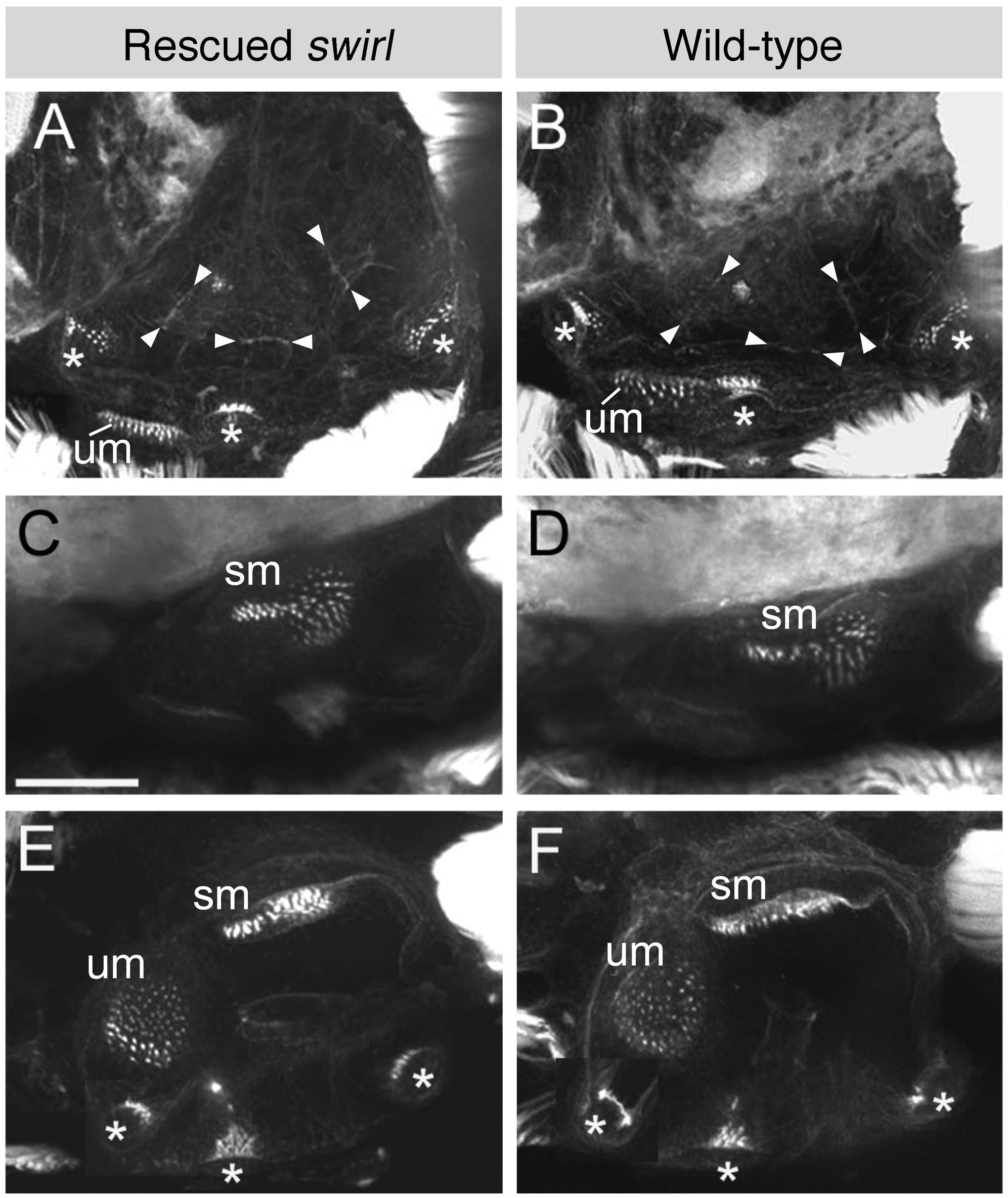
Fig. 3 Sensory patches and fusion plates are present in the inner ears of 5 dpf rescued swr embryos.
Projected confocal z-stacks through inner ears of 5 dpf embryos stained with FITC-conjugated phalloidin, revealing the actin-rich stereociliary bundles of the sensory hair cells and the cortical actin in every cell. (A–D) Lateral views; anterior to left, dorsal to top. (A, B) Lateral plane of focus, showing the cristae, utricular macula and the fusion plates (arrowheads) between the semicircular canal projections. (C, D) More medial plane of focus showing the saccular macula. (E, F) Dorsal views; anterior to left, medial to top. Images shown are composites of two sets of projected z-stacks as the anterior crista is in a more dorsal plane of focus than the remaining sensory patches. Abbreviations: sm, saccular macula; um, utricular macula. Cristae are indicated with an asterisk. Scale bar, 50 μm.