
|
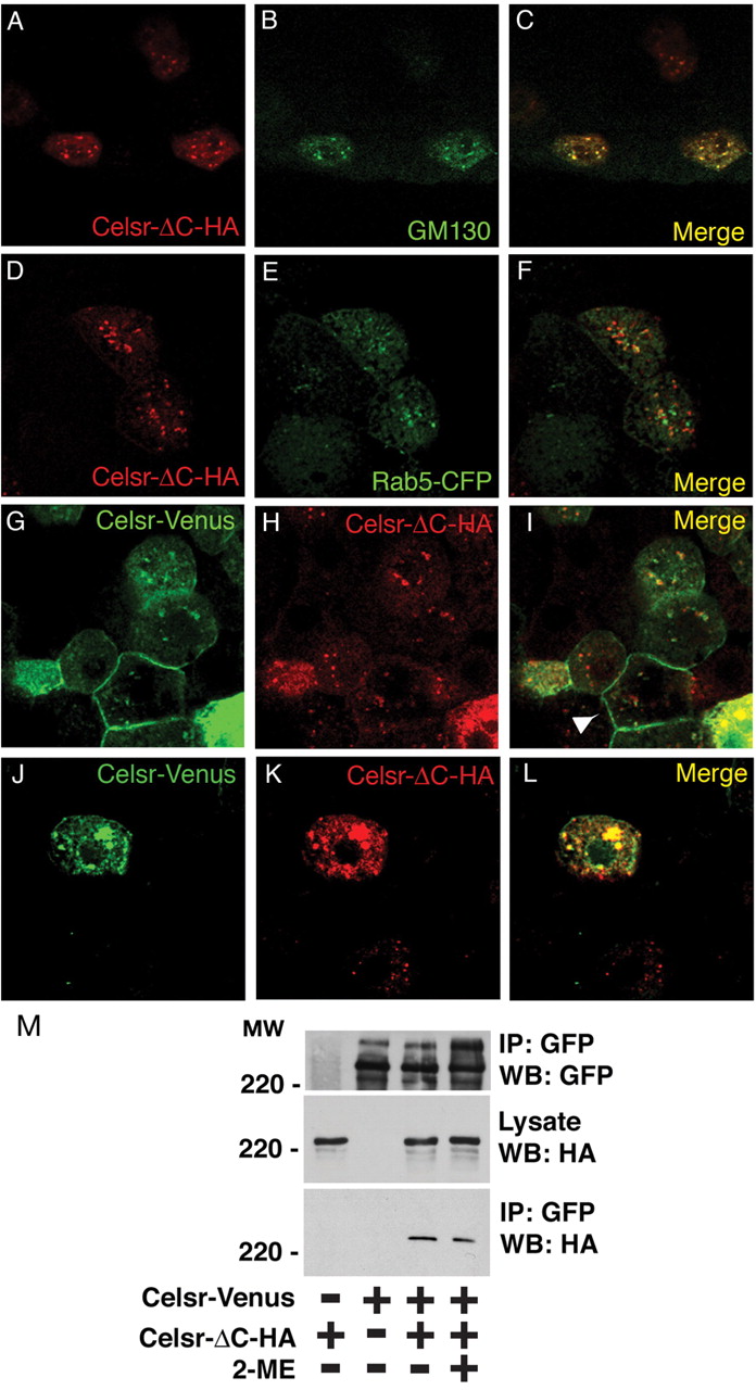
Fig. 4 Celsr-ΔC is retained in the golgi and inhibits membrane presentation of Celsr. (A-F) Wild-type embryos were injected with 30 pg Celsr-ΔC-HA DNA alone (A-C) or together with 30 pg Rab5-CFP RNA (D-F) and fixed at 40% epiboly to visualise with an HA antibody (A,D) or either a GM130 antibody for the golgi (B) or a GFP antibody for early endosomes (E). The merge images are shown in C and F. (G-L) Wild-type embryos were co-injected with 30 pg Celsr-Venus DNA and 30 pg Celsr-ΔC-HA DNA, and fixed at 40% epiboly to visualise with a GFP antibody (G,J) or an HA antibody (H,K). The merge images are shown in I and L. When the level of Celsr-ΔC is low, Celsr-Venus is presented at the membrane (shown by arrowhead) (62 cells out of 15 embryos examined but the level of Celsr-ΔC is high, Celsr-Venus is prevented from presenting at the membrane (15 cells out of 15 embryos examined). (M) Non-covalent dimer formation of Celsr. Celsr-Venus and/or Celsr-deltaC-HA were transiently expressed in HEK293 cells. Cell lysates were immunoprecipitated with anti-GFP antibody, and immunoprecipitated proteins were further incubated in Laemmli's buffer with or without 2-mercaptoethanol (2-ME), followed by western blotting with anti-GFP or anti-HA antibody.