
|
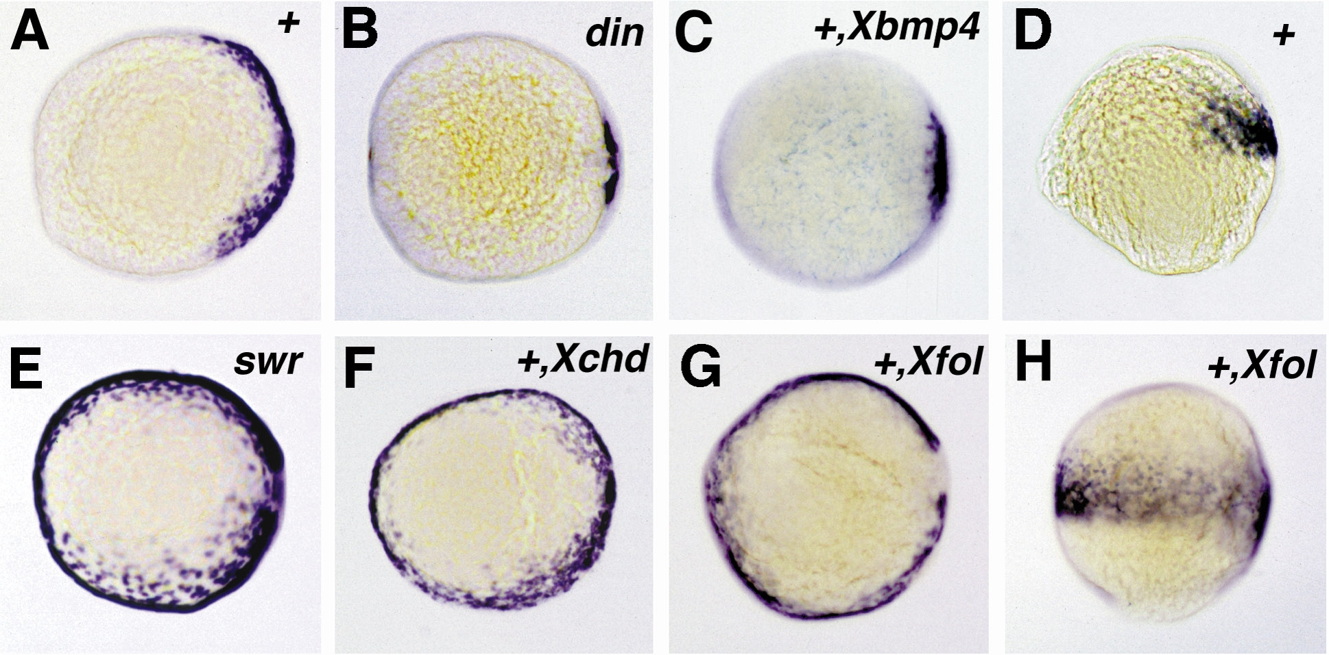
Fig. 6 Expression of follistatin in chordino and bmp2b mutants and after overexpression of Xenopus follistatin,bmp4, or chordin. All embryos shown are at 90% epiboly and are viewed from the animal pole, except in (D and H), which are lateral views. Expression of follistatin is reduced in the chordino mutant embryo (B), spanning the dorsalmost 30° of the embryonic circumference, compared to 150° in wild-type sibling embryos (A). A similar dorsal retraction of the follistatin expression domain is obtained in wild-type embryos after injection of synthetic Xenopus bmp4 mRNA (C). In the bmp2b mutant embryo (E), the follistatin expression domain is expanded, spanning the entire embryonic circumference. The same effect is achieved in wild-type embryos by the injection of Xenopus chordin (F) and Xenopus follistatin mRNA (G). In (H), the Xfol-injected embryo of (G) is shown in a lateral view in comparison to an uninjected wild-type sibling embryo (D), showing that ectopic ventral zebrafish follistatin expression is restricted to the same anteroposterior levels like the wild-type expression on the dorsal side. Abbreviations: +, wildtype; din, chordino mutant dintt250; swr, bmp2b mutant swrta72. The second abbreviation in the upper right corner indicates the injected mRNA: Xbmp4, Xenopus bmp4; Xchd, Xenopus chordin; Xfol, Xenopus follistatin.
Reprinted from Developmental Biology, 204, Bauer, H., Meier, A., Hild, M., Stachel, S., Economides, A., Hazelett, D., Harland, R.M., and Hammerschmidt, M., Follistatin and Noggin are excluded from the zebrafish organizer, 488-507, Copyright (1998) with permission from Elsevier. Full text @ Dev. Biol.