
|
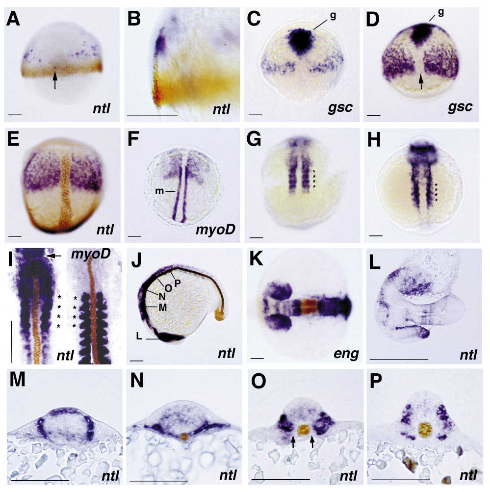
Fig. 5 Expression pattern of follistatin in wild-type zebrafish embryos, revealed by whole-mount in situ hybridization. All embryos except the right embryo in (I) are stained for follistatin transcripts. In the case of double in situ hybridization immunostainings (A, B, E, I–K, M–P) and double in situ hybridizations (C, D, F), the second detected gene product is indicated in the lower right corner. (A and B) 60% epiboly, double stained for No tail protein (brown), dorsal view (A) and close-up of same embryo in dorsolateral view (B); scattered follistatin expression is seen in anterior paraxial regions and occasionally in single cells (indicated by an arrow, also in D) in posterior axial positions within the no tail expression domain in the notochord anlage (A). Expression of follistatin is restricted to involuted cells of the hypoblast (B). (C, D, and E) 80% epiboly (C), 90% epiboly (D), and tailbud stage (E), double stained for goosecoid (gsc) RNA (C, D, indicated with g) and No tail protein (E, brown), dorsal view; the paraxial follistatin expression domain grows, but expression remains excluded from anterior axial regions (E) which are characterized by the expression of the organizer-specific gene goosecoid (C, D). Posterior axial follistatin staining is weaker than staining in paraxial regions. In addition, the intensity of the axial follistatin staining varies in specimens from different in situ hybridizations (compare, e.g., C, D, and Fig. 7B). The reason for this variability is currently unknown. (F) 2-somite stage, double stained for myoD RNA (m), dorsal view; the posterior region of the follistatin expression domain and the anterior region of the myoD expression domain overlap, indicating that follistatin is expressed in the presumptive head mesoderm and a distinct anterior region of the presomitic mesoderm. The follistatin expression in the anterior presomitic mesoderm appears slightly condensed. (G and H) 5-somite stage (G) and 10-somite stage (H), dorsal view; the posterior paraxial follistatin expression is organized in a segmented pattern defining four stripes (indicated by asterisks) at identical anteroposterior positions in embryos of both stages. No additional stripes are visible in the 10-somite embryo. Note the intense anterior staining in (H), representing expression in the brain anlage. (I) 12-somite stage, stained for follistatin RNA and No tail protein (left) and for myoD RNA and No tail protein (right), dorsal view on spread embryos. In the left embryo, the anterior tip of the notochord anlage is indicated by an arrow. Comparison of the follistatin and myoD expression patterns assigns the intense segmented follistatin expression to the first 4 somites (indicated by asterisks). In addition, weaker follistatin expression is visible in somites 5 to 7. In all somites, follistatin expression is excluded from adaxial regions. (J) 20-somite stage, double stained for No tail protein, lateral view. Intense follistatin staining is observed in the head and anterior trunk region, weaker staining in more posterior somites. Indicated are the positions for which sections of similar embryos are shown in (L–P). (K) 20-somite stage, double stained for Engrailed protein; neuroectodermal follistatin expression is confined to the eye vesicles and four distinct bands in the forebrain, midbrain, and hindbrain, while the midbrain–hindbrain boundary region, which is characterized by the expression of engrailed, lacks follistatin transcripts. (L–O) Saggital (L) and transverse (M–P) 7-μm paraffin sections of doubly stained embryos of the 16-somite (M, L) and 25-somite (O, N, P) stage at the respective positions indicated in (J). (L) Section through the ventral (upper eye) and the medial (lower eye) part of the eye vesicles, revealing follistatin expression in posterior and distal regions of the eye primordia. (M) At the level of the anterior hindbrain, paraxial follistatin expression is restricted to an approximately three cell diameter broad region surrounding the neural tube. In the brain, follistatin is expressed in a distinct dorsal band and a more diffuse band in the ventral part of the tube. (N) More posteriorly, in the notochord region anterior of the somites, follistatin is expressed in the narrow band of cells located between the neural tube and the yolk and in ventral paraxial cells. (O) In the first 4, relatively small somites, follistatin is expressed throughout the entire somitic region except the adaxial positions adjacent to the notochord. Note the weak diffuse follistatin expression in the neural tube. (P) In posterior somites, follistatin expression is confined to ventral regions, which most likely represent the presumptive sclerotome, and to dorsal regions of the myotome in the vicinity of the neural tube. Abbreviations: eng, Engrailed; g, gsc,goosecoid; m, myoD, ntl, No tail.
Reprinted from Developmental Biology, 204, Bauer, H., Meier, A., Hild, M., Stachel, S., Economides, A., Hazelett, D., Harland, R.M., and Hammerschmidt, M., Follistatin and Noggin are excluded from the zebrafish organizer, 488-507, Copyright (1998) with permission from Elsevier. Full text @ Dev. Biol.