
|
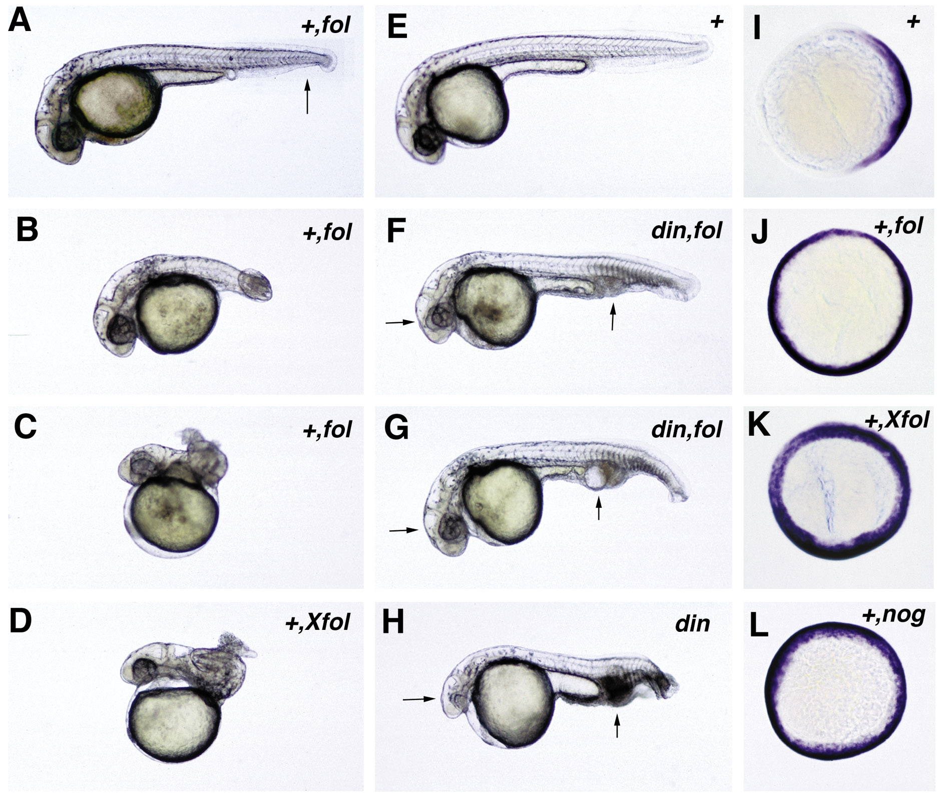
Fig. 2 Functional analysis of zebrafish follistatin and noggin. Both molecules can dorsalize zebrafish embryos. (A–D) Overexpression of zebrafish and Xenopus follistatin in wild-type embryos leads to a dose-dependent dorsalization. Weakly dorsalized embryos (C1) display a partial loss of the ventral tail fin, as indicated in (A) by an arrow. Moderate dorsalization (C3) is characterized by a wound-up tail, the “piggy tail” (Mullins et al., 1996) phenotype (B), while in strongly dorsalized embryos (C4/C5), the entire embryonic axis is wound up in a snailshell-like fashion (C, D). (E–H) chordino mutant embryos can be rescued by overexpression of zebrafish follistatin (F, G). Note the normalization of eye sizes and blood islands (indicated by arrows in F–H). All embryos in (A–H) are shown at approximately 36 hpf. (I–L) Expression of fkd3; 70% epiboly, animal view, dorsal right; in the wild-type embryo. (I) fkd3 expression is confined to the dorsal side, while it is expanded into ventralmost regions in embryos injected with zebrafish follistatin (J), Xenopus follistatin (K), or zebrafish noggin (L). Abbreviations: +, wildtype; hpf, hours after fertilization; din,chordino mutant dintt250. The second abbreviation in the upper right corner indicates the injected mRNA: fol, zebrafish follistatin; Xfol, Xenopus follistatin; nog, zebrafish noggin.
Reprinted from Developmental Biology, 204, Bauer, H., Meier, A., Hild, M., Stachel, S., Economides, A., Hazelett, D., Harland, R.M., and Hammerschmidt, M., Follistatin and Noggin are excluded from the zebrafish organizer, 488-507, Copyright (1998) with permission from Elsevier. Full text @ Dev. Biol.