
|
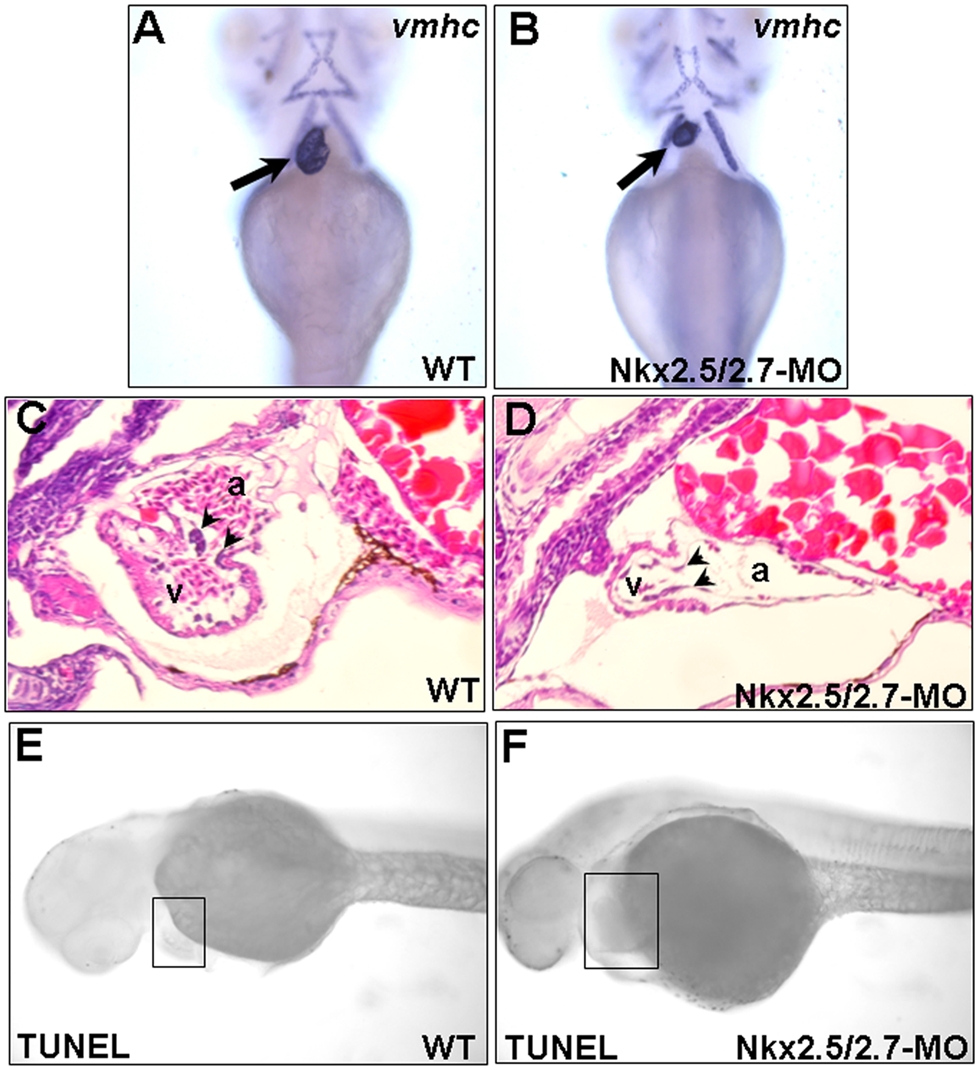
Fig. 3 Ventricle becomes smaller and consists of a single layer in the Nkx2.5 and Nkx2.7 double knockdown morphants (Nkx2.5/2.7-MO).
Ventricular myosin heavy chain (vmhc) was used as a probe to detect the ventricle morphology in (A) wild-type (WT) and (B) Nkx2.5/2.7-MO embryos at 72 hpf. The ventricle of Nkx2.5/2.7-MO embryos was smaller than that of WT embryos (indicated by arrows). Hematoxylin and eosin staining showed that ventricular myocardium of WT was two or more cell layers in thickness. However, only one cell layer was retained in the ventricular myocardium of the Nkx2.5/2.7-MO embryos. In addition, compared to wild-type embryos, the endocardium of Nkx2.5/2.7-MO-injected embryos did not form endocardial cushion (indicated by arrowheads in C and D). Like WT embryos, TUNEL assay did not display the increase of TUNEL-positive cells in the heart region of Nkx2.5/2.7-MO embryos at 40 hpf (indicated by boxes in E and F). Embryos were observed ventrally (A–B) or laterally (C–F). v: ventricle; a: atrium.