
|
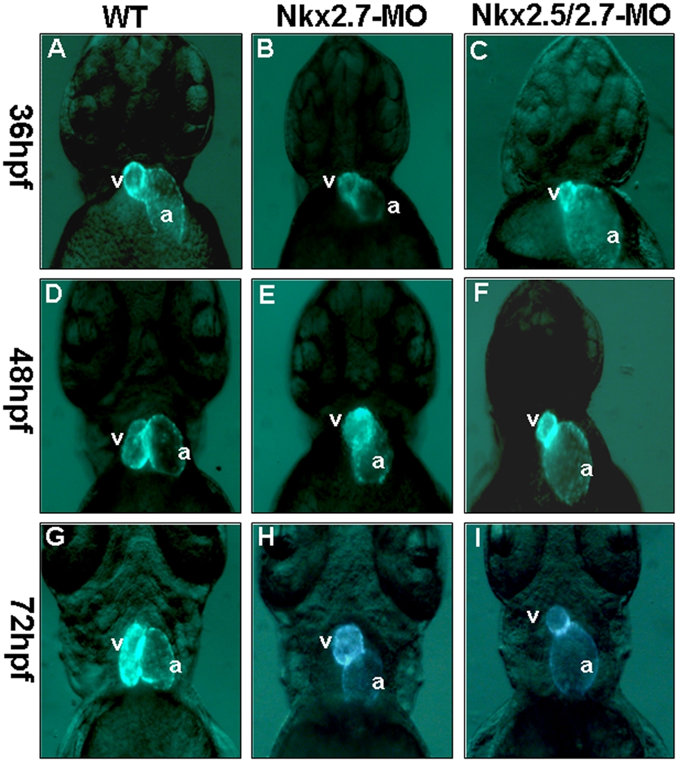
Fig. 1 The defective phenotypes of zebrafish embryo heart injected with Nkx2.5-MO, Nkx2.7-MO and Nkx2.5/2.7-MO.
Eight nanograms of MO were injected into one-cell stage embryos derived from transgenic line Tg (cmlc2::GFP) to knock down the Nkx protein specifically. The embryos are shown at 36 hpf (A, B, C), 48 hpf (D, E, F), and 72 hpf (G, H, I). The heart phenotype of Nkx2.5-MO embryos was similar to that of control embryos whose ventricle is located at the right side of the atrium when embryos were observed at 36 hpf, 48 hpf and 72 hpf from the ventral view under fluorescence microscope (A, D, G). However, embryos injected with Nkx2.7-MO displayed an unlooping defect from 36 hpf to 72 hpf (B, E, H). Embryos injected with Nkx2.5/2.7-MO displayed a shrunken ventricle and an expanding atrium (C, F, I). v: ventricle; a: atrium.