
|
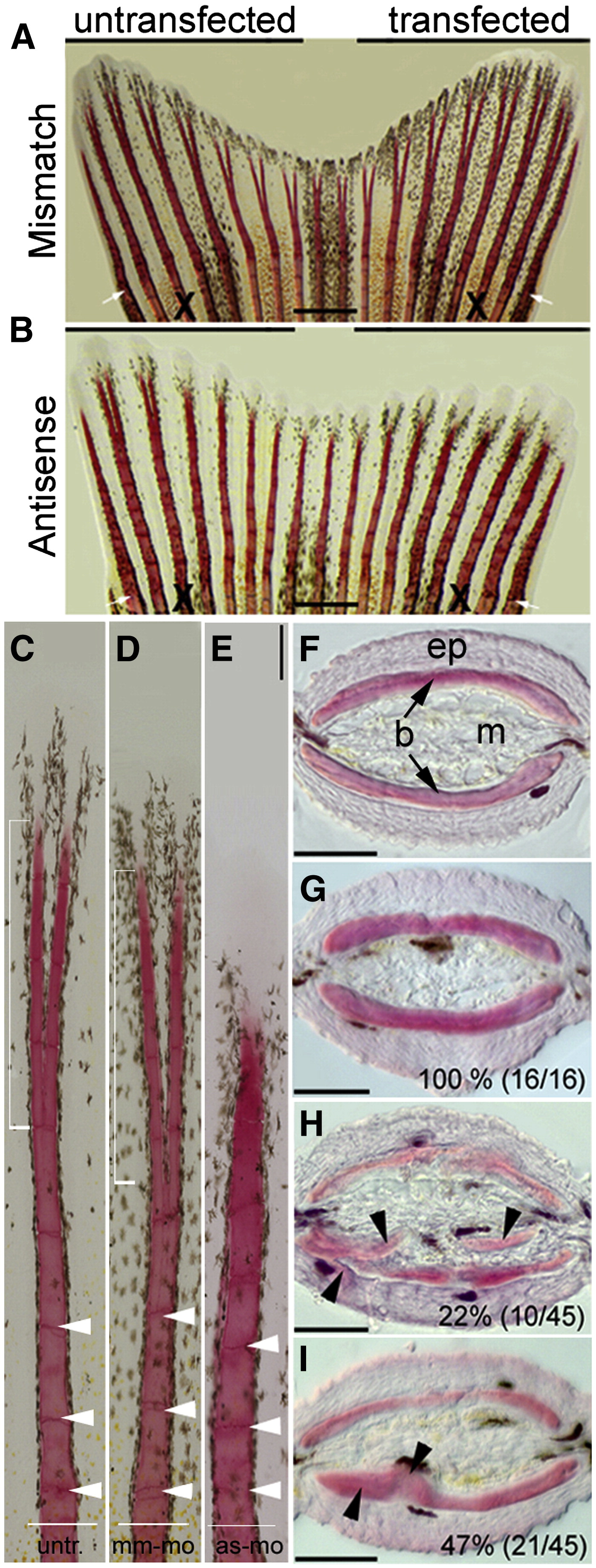
Fig. 7 Knockdown of smp results in ectopic bone formation in regenerating fins. (A) Zebrafish caudal fins were allowed to regenerate 5 days after transfecting one lobe with mismatch morpholino (7 dpa). (B) Fin lobes transfected with antisense morpholino. White arrow indicate amputation plane and “X” indicates the compared rays. (C) Individual rays from untransfected lobes show regular segmentation (white arrow heads) and distal bifurcation of the ray (white square bracket). (D) Rays from mismatch-morpholino-transfected fin. (E) Bony rays from antisense-morpholino-transfected fin lobes with shorter and thicker bone segments (white arrowheads). Distal bifurcation is not visible. (F) Axial cross sections through fin show the epidermis (ep), the two hemi-rays bones (b) and interray mesenchyme (m) from one lepidotrichia. (G) Cross sections through hemi-rays of mismatch-morpholino-transfected fin lobe. Inset number indicates phenotype/total (H) Cross section through hemi-ray of antisense-morpholino-transfected fin lobe shows ectopic bone (black arrow heads). (I) Antisense-morpholino-transfected fins displayed regional thickening within the fin bones (black arrow heads). Scale bars equal 500 μm (A and B) and 100 μm (C–I).
Reprinted from Developmental Biology, 325(2), Kizil, C., Otto, G.W., Geisler, R., Nüsslein-Volhard, C., and Antos, C.L., Simplet controls cell proliferation and gene transcription during zebrafish caudal fin regeneration, 329-340, Copyright (2009) with permission from Elsevier. Full text @ Dev. Biol.