
|
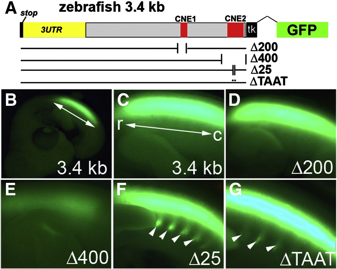
Fig. S3 Deletions in the zebrafish evx1 enhancer cause de-regulation of reporter GFP expression in the chick. Embryos were electroporated with deletion variants of 3.4 kb::tkGFP (from Fig. 4A). Δ200 removes CNE1, Δ400 removes CNE2, Δ25 removes 25HCS and ΔTAAT is a double deletion of two homeodomain (TAAT) sites in 25HCS. (B) Low magnification image of GFP in a control (3.4 kb) whole-mount chick embryo. Bright GFP signal is observed along the rostral (r) to caudal (c) axis of the trunk (double arrow in B,C) within the spinal cord. (C–G) High magnification images focusing on a side view of the spinal cord. (C) control 3.4 kb. (D) Δ200 is indistinguishable from the control. (E) Δ400 dramatically eliminated GFP expression. (F, G) Δ25 and ΔTAAT ectopically labeled peripheral motor axon bundles that exit the spinal cord (arrowheads).
Reprinted from Developmental Biology, 325(2), Suster, M.L., Kania, A., Liao, M., Asakawa, K., Charron, F., Kawakami, K., and Drapeau, P., A novel conserved evx1 enhancer links spinal interneuron morphology and cis-regulation from fish to mammals, 422-433, Copyright (2009) with permission from Elsevier. Full text @ Dev. Biol.