
|
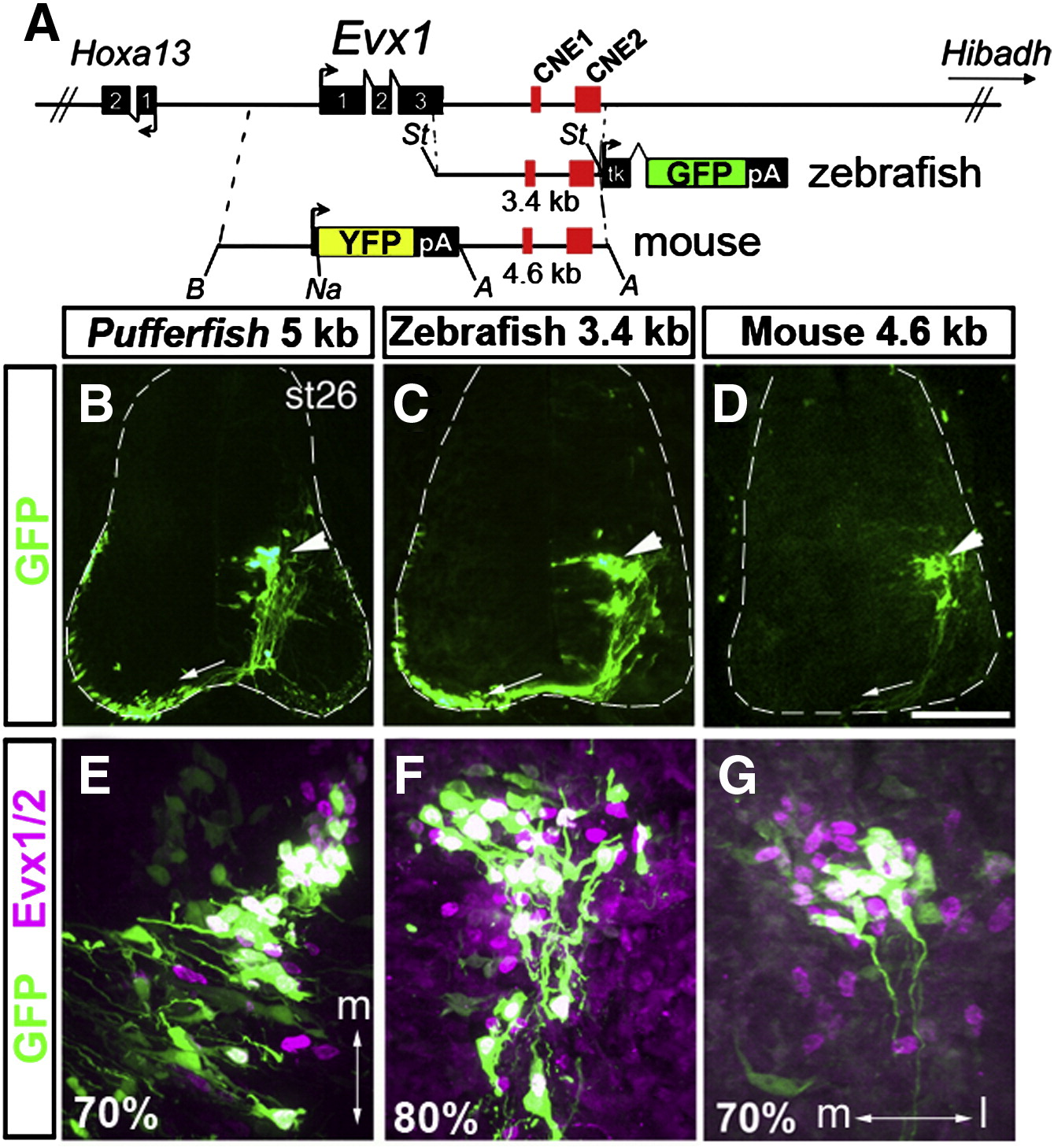
Fig. 4 Zebrafish and mouse Evx1 downstream fragments direct gene expression to chick V0 interneurons. (A) Schematic of the Evx1 genomic region and the fragments used to generate the constructs for electroporation in the chick. A 3.4 kb zebrafish fragment including the last 89 bp of exon 3, 3′ UTR and CNE1 and CNE2 was placed upstream of the tkGFP cassette. The mouse reporter construct consists of 3.6 kb upstream and 4.6 kb downstream. YFP was fused in frame to exon 1 (+ 156 from ATG start) in the latter. St, StuI, B, BamHI, Na, NaeI and A, ApaI. (B–G) Representative sections from electroporated embryos. Pufferfish (B), zebrafish (C) and mouse (D) constructs show a highly similar pattern of GFP or YFP expression localized to a cluster of commissural interneurons (arrowheads) whose axons can be seen crossing to the opposite side of the cord (arrows). The outline of the spinal cord is drawn as dashed lines. Scale bar: 110 μm. (E,F,G) Magnified views of GFP+ cells double labeled with Evx1/Evx2 antibodies. Medial (m) and lateral (l) orientation is indicated by the double arrow and is the same for F and G. Percentage of GFP+ cells that co-expressed Evx1/Evx2 on average is indicated at the bottom left corner of each panel (see text for details). At least 6 embryos were examined for each construct.
Reprinted from Developmental Biology, 325(2), Suster, M.L., Kania, A., Liao, M., Asakawa, K., Charron, F., Kawakami, K., and Drapeau, P., A novel conserved evx1 enhancer links spinal interneuron morphology and cis-regulation from fish to mammals, 422-433, Copyright (2009) with permission from Elsevier. Full text @ Dev. Biol.