
Image
|
Figure Caption
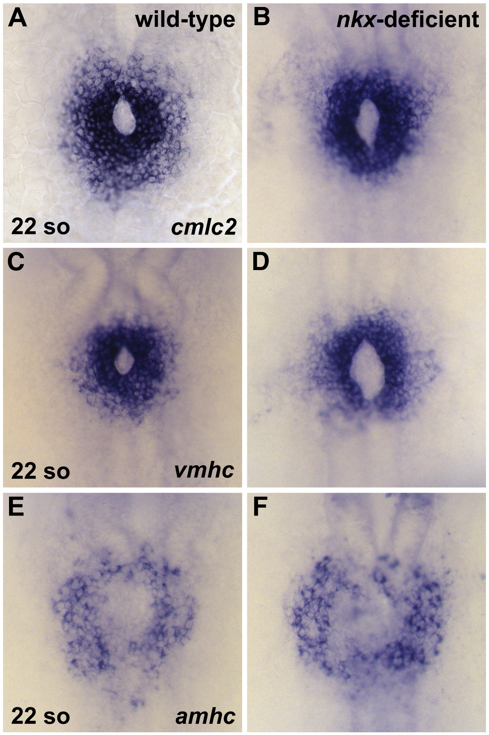
Fig. 4 Cardiac cone formation occurs normally in nkx-deficient embryos. In situ hybridization depicts expression of cmlc2 (A,B), vmhc (C,D), and amhc (E,F) in wild-type and nkx-deficient embryos. Embryos viewed dorsally, anterior to the top, at the 22-somite stage. (A,C,E) Wild-type embryos exhibit normal fusion of the bilateral cardiac precursors, creating an intact cardiac cone. (B,D,F) nkx-deficient embryos mirror the expression patterns observed in wild-type, with the exception of a slightly enlarged and elongated lumen within the cardiac cone.
Figure Data
Acknowledgments
This image is the copyrighted work of the attributed author or publisher, and
ZFIN has permission only to display this image to its users.
Additional permissions should be obtained from the applicable author or publisher of the image.
Reprinted from Developmental Biology, 322(2), Targoff, K.L., Schell, T., and Yelon, D., Nkx genes regulate heart tube extension and exert differential effects on ventricular and atrial cell number, 314-321, Copyright (2008) with permission from Elsevier. Full text @ Dev. Biol.