
|
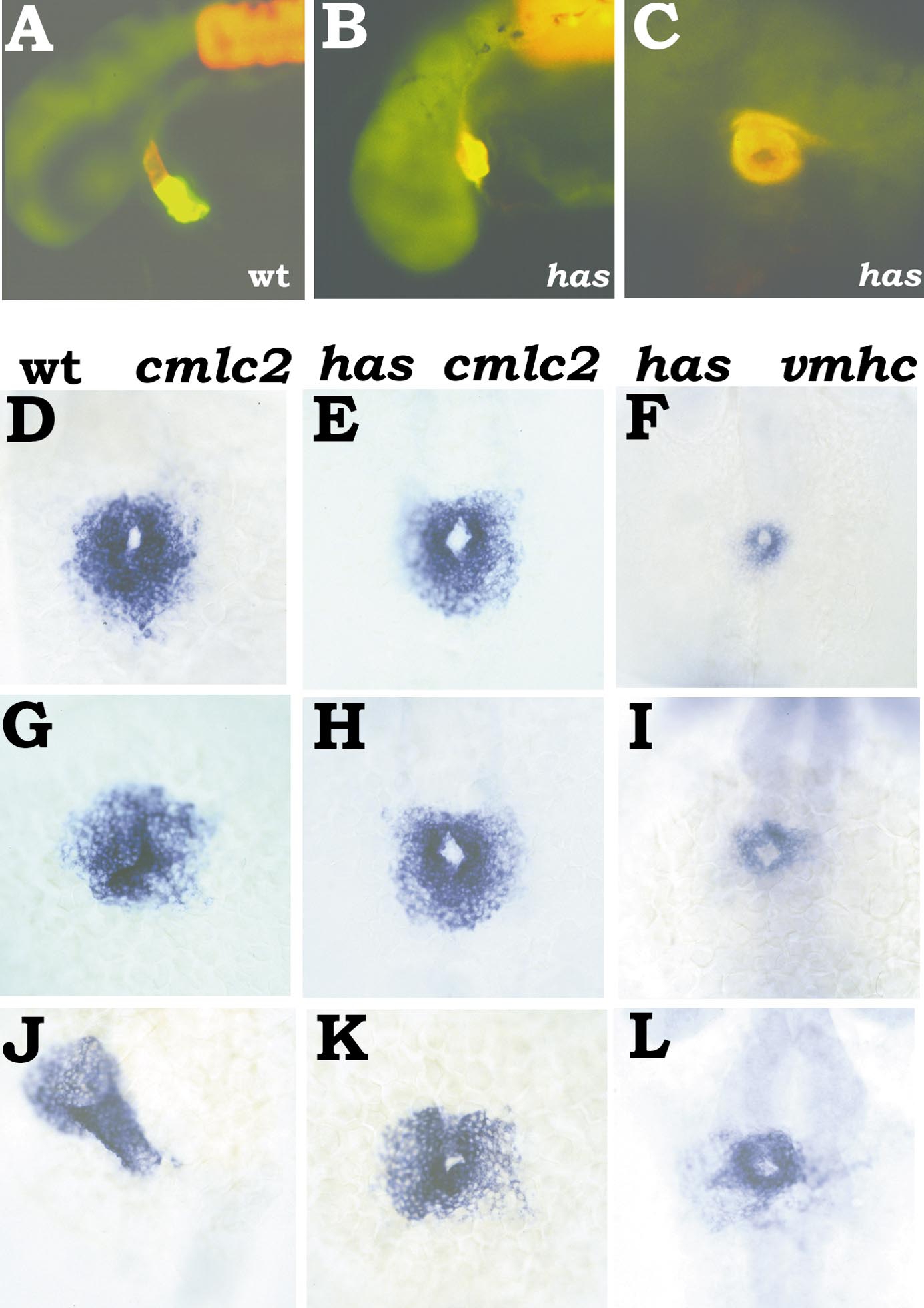
Fig. 6 heart and soul (has) disrupts heart tube assembly at an intermediate stage. (A, B, C) 27-hpf embryos stained with MF20 (TRITC) and S46 (FITC), anterior to the left. Red fluorescence indicates MF20 staining of ventricular tissue, while yellow fluorescence indicates the overlap of S46 and MF20 staining in atrial tissue. (A) Lateral view of wild-type embryo. The atrium (yellow) lies posterior to the ventricle (red). (B) Lateral view of has mutant. (C) Ventral view of has mutant. The atrium (yellow) surrounds the ventricle (red). (D–L) Dorsal views, anterior at the top, of wild-type and has mutant embryos. (D, G, J) Expression of cmlc2 in wild-type embryos. (E, H, K) Expression of cmlc2 in has mutant embryos. (F, I, L) Expression of vmhc in has mutant embryos. (D, E, F) 21-somite stage; the cardiac cone has formed and has mutants are indistiguishable from their wild-type siblings. (G, H, I) 23-somite stage; while the apex of the cone has tilted in wild-type embryos (G), the cone in has mutants remains stationary (H, I). (J, K, L) 24 hpf; the has heart still retains a cone-like structure (K, L), even as formation of the wild-type heart tube is nearly complete (J).
Reprinted from Developmental Biology, 214(1), Yelon, D., Horne, S.A., and Stainier, D.Y.R., Restricted expression of cardiac myosin genes reveals regulated aspects of heart tube assembly in zebrafish, 23-37, Copyright (1999) with permission from Elsevier. Full text @ Dev. Biol.