
|
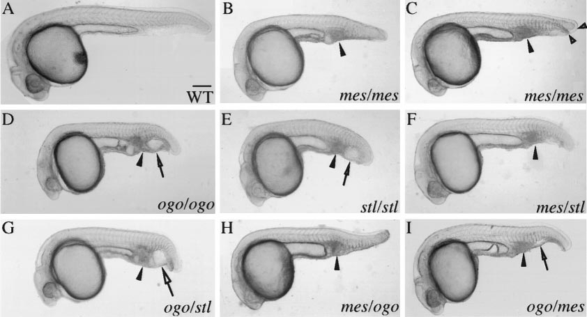
Fig. 1 mes, ogo, and stl fail to complement. (A) WT (1 day), (B,C) mes, (D) ogo, and (E) stl homozygotes (1 day) are typically characterized by a shortened body axis, an accumulation of cells in the ventral tail (arrowhead), and a partial duplication of the ventral fin. In addition, ogo and stl homozygotes have smaller heads and an enlarged vesicle in the ventral tail (arrow). (B) Progeny of a mes/+ female. (C) Progeny of mes/mes females exhibit a more prominent duplicated ventral fin (open arrowheads). (F–I) mes, ogo, and stl mutations fail to complement. All matings produced transheterozygous mutant progeny (Table 1). (H and I) A maternal enhancement of mutant phenotypes was revealed in reciprocal crosses. The enlarged tail vesicle (open arrow) was not present in the (H) ogo/mes progeny of a mes/+ female, but was observed in (I) progeny of ogo/+ females. The phenotype of ogo/mes transheterozygotes was of intermediate severity compared to (C) mes and (D) ogo homozygotes, suggesting that mes is a hypomorphic allele. The maternal allele is given first in the genotype designations. Scale bar, 200 μm for (A–I).
Reprinted from Developmental Biology, 214(1), Miller-Bertoglio, V., Carmany-Rampey, A., Fürthauer, M., Gonzalez, E.M., Thisse, C., Thisse, B., Halpern, M.E., and Solnica-Krezel, L., Maternal and zygotic activity of the zebrafish ogon locus antagonizes BMP signaling, 72-86, Copyright (1999) with permission from Elsevier. Full text @ Dev. Biol.