
Image
|
Figure Caption
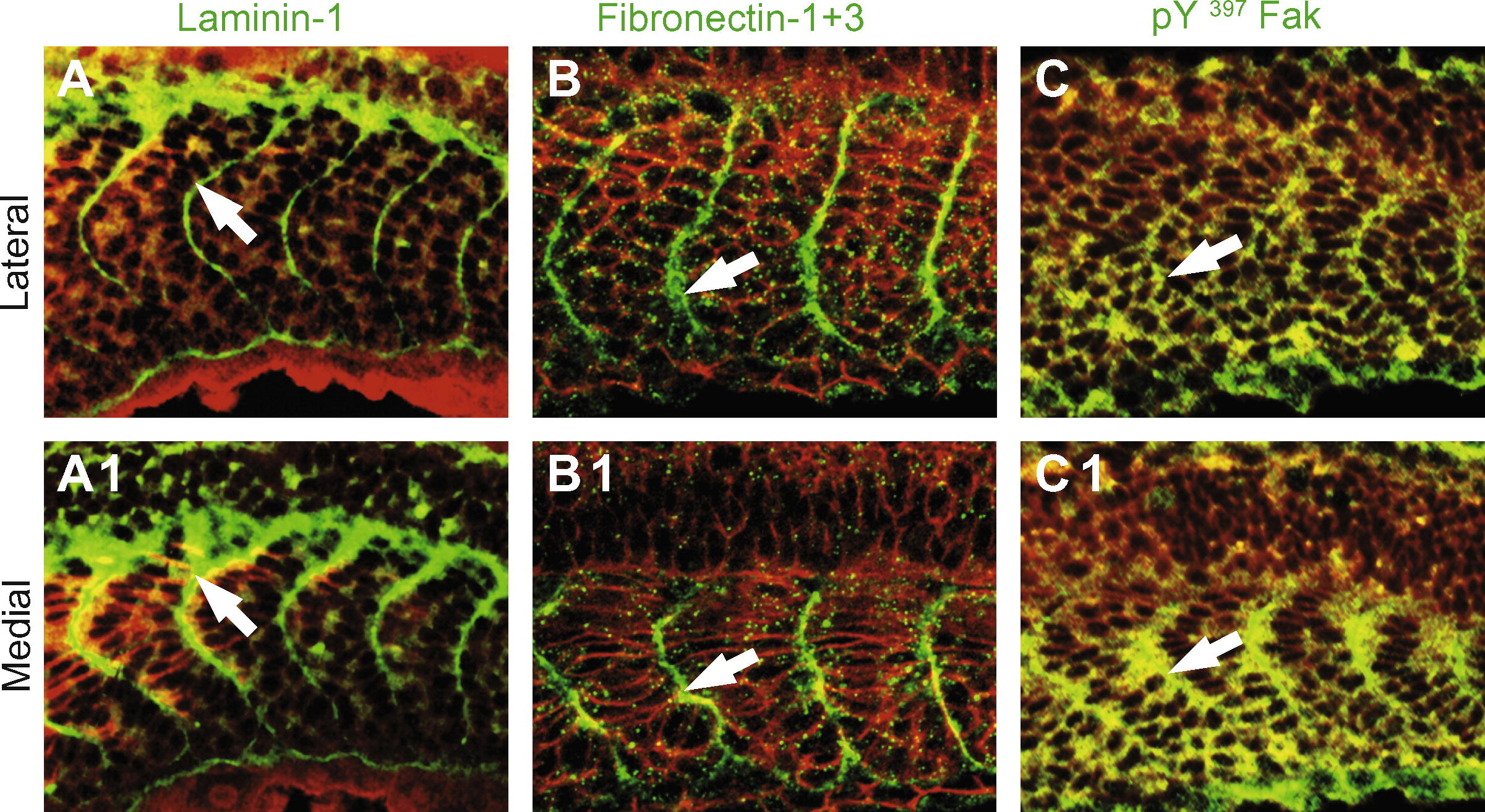
Fig. 1 The ECM proteins laminin-1, Fn and Fak concentrate at the initial epithelial somite boundary. All panels are Leica confocal micrographs. All panels side view, anterior left, dorsal top, of 18-somite wild-type embryos. Panels (A–C) are superficial views and panels numbered 1 are medial views of the same Z-stack. β-Catenin which outlines all cells is in red, and laminin, Fn and Fak are each in green. Laminin-1 (A, A1, white arrows), Fn (B, B1, white arrows), and pY397 FAK (C, C1, white arrows) are all concentrated at the somite boundaries throughout the medial-lateral axis of the embryo.
Figure Data
Acknowledgments
This image is the copyrighted work of the attributed author or publisher, and
ZFIN has permission only to display this image to its users.
Additional permissions should be obtained from the applicable author or publisher of the image.
Reprinted from Gene expression patterns : GEP, 9(1), Snow, C.J., and Henry, C.A., Dynamic formation of microenvironments at the myotendinous junction correlates with muscle fiber morphogenesis in zebrafish, 37-42, Copyright (2009) with permission from Elsevier. Full text @ Gene Expr. Patterns