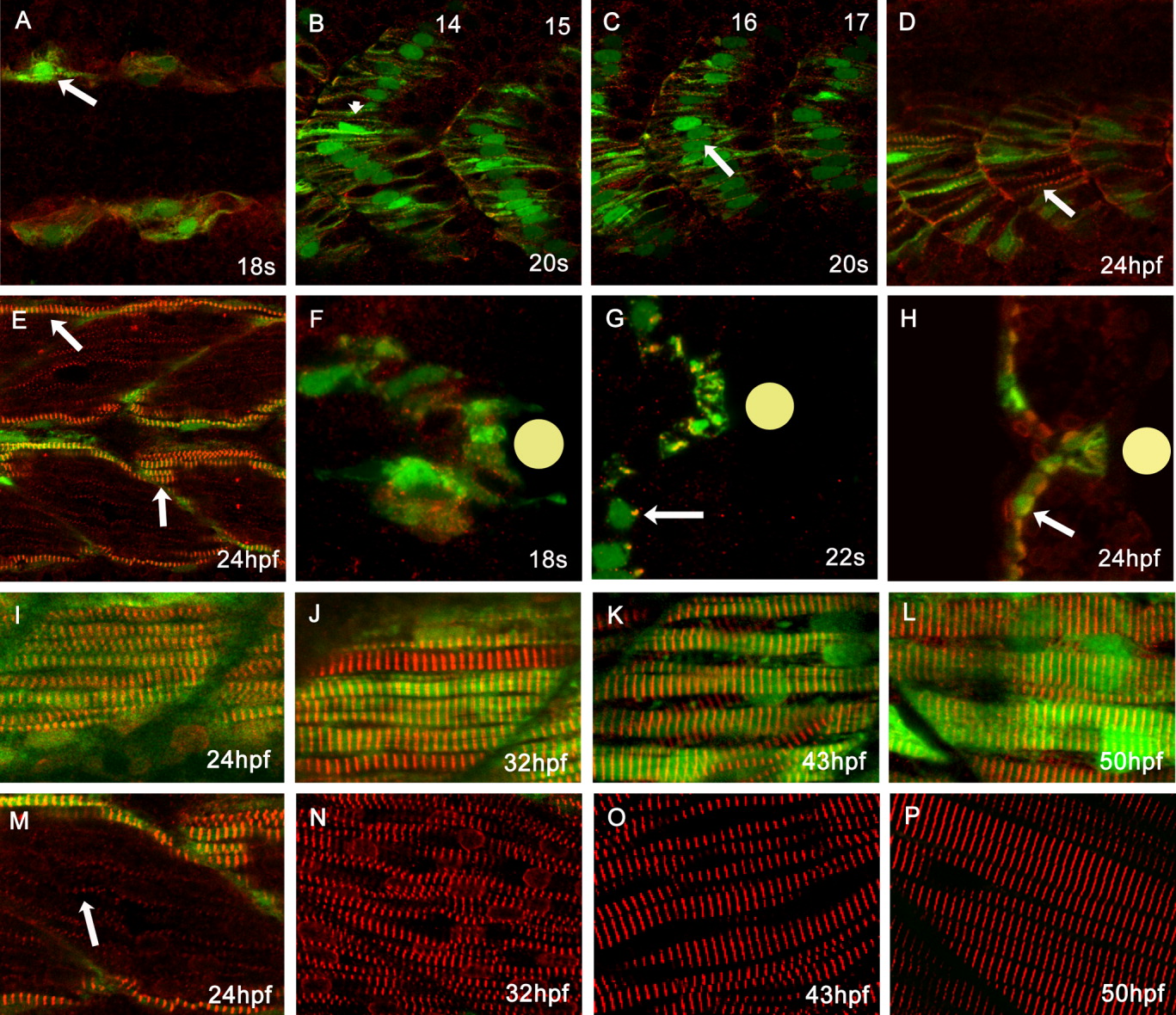
Fig. 6 Normal myofibrillogenesis in Tg(smyhc1:GFP)i104 transgenic embryos. A: Dorsal view of an 18 somite stage embryo showing the first appearance of striated α-Actinin (red: arrow) in slow muscle fibres (marked by GFP expression). B,C: Lateral aspect of a 20-hpf embryo: α-Actinin was organised as a clear z-band (arrowhead) in anterior somites (B), but not in more posterior somites (C, arrow). D,E: Lateral aspect of 24-hpf embryo: posterior slow fibres have clearly discernible striated thin myofibrils (D) while in more anterior somites the myofibrils have become quite thick (E, arrows). F,G: Transverse sections at successive stages reveal that in slow fibre progenitors, prior to their migration away from the notochord (indicated by the yellow disc) α-Actinin distribution is quite diffuse (F), but as migration proceeds, it becomes localised to one side of the nucleus (G, arrow) and 24 hpf reveals the fully assembled myofibrils (H). I-P: Pairs of images of lateral aspects of the surface slow muscle and the underlying fast muscle at successively later stages of development: fast muscle myotubes started to express and localise α-Actinin as a single file of myofibrils at 24 hpf, much later than in slow muscle fibres (M: arrow). Myofibrillogenesis proceeds rapidly in both slow and fast lineages between 32-50 hpf.
Image
Figure Caption
Figure Data
Acknowledgments
This image is the copyrighted work of the attributed author or publisher, and
ZFIN has permission only to display this image to its users.
Additional permissions should be obtained from the applicable author or publisher of the image.
Full text @ Dev. Dyn.

