
Image
|
Figure Caption
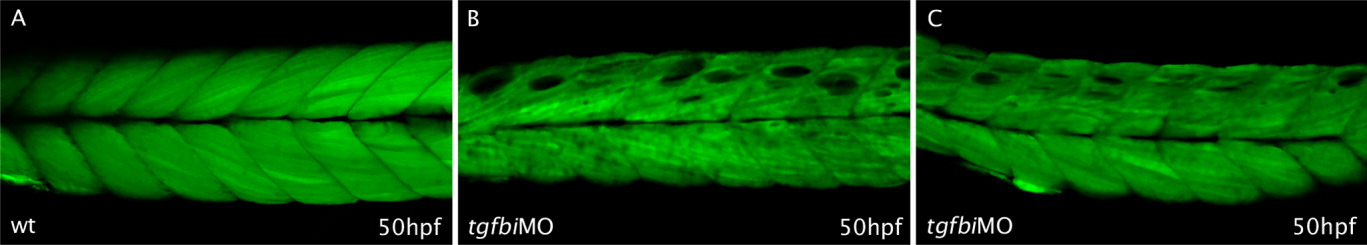
Fig. 4 Loss of Tgfbi causes lateral detachment of muscle fibres. Live images of Tg(acta1:GFP)zf13 50-hpf control and morphant embryos. A: In wild type embryos, the muscle fibres are tightly connected to each other along their length. B: In tgfbi start site morphant embryos, by contrast, empty gaps were apparent in between muscle fibres. C: The same phenotype was observed following injection of a mixture of the start and splice morpholinos, each at half the concentration of that effective for single morpholinos.
Figure Data
Acknowledgments
This image is the copyrighted work of the attributed author or publisher, and
ZFIN has permission only to display this image to its users.
Additional permissions should be obtained from the applicable author or publisher of the image.
Full text @ Dev. Dyn.