
|
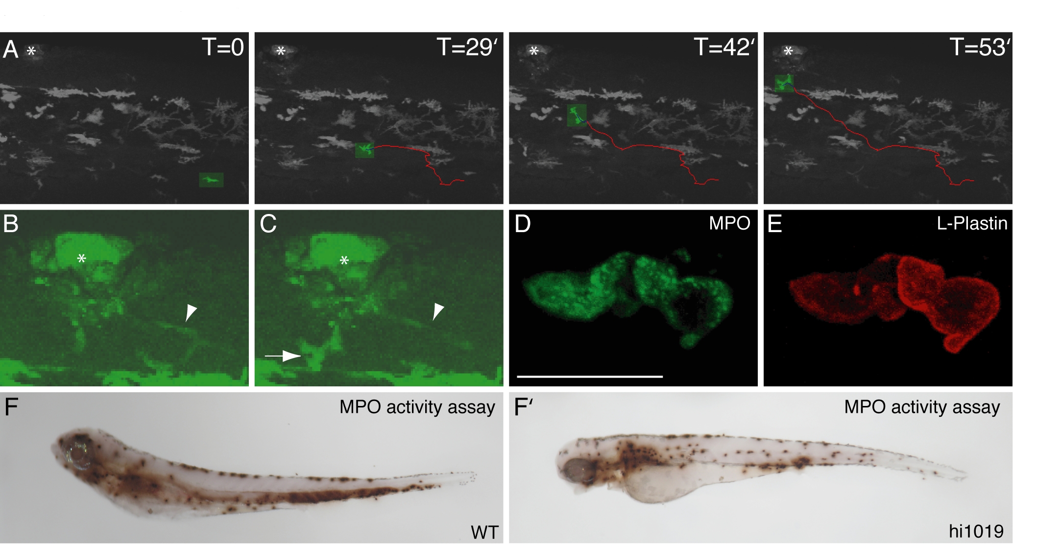
Fig. S2 Neutrophils in fad24hi1019 larvae respond to wounds and have active MPO contained in cytoplasmic granules. A: A fad24hi1019;mpx:GFP mutant at 3 dpf was analyzed by time-lapse confocal microscopy. Frames are taken from Supp. Movie 3. The asterisk indicates a wound made in the dorsal fin. A single neutrophil is marked in each frame in green with a red track indicating the path it takes from the body to the fin. Indicated times are from the start of the movie. B: A blown-up image of the wound (taken from Supp. Movie 4) reveals the presence of a low-level MPO-expressing cell with a long and extended morphology (arrowhead). C: Both a low-level MPO-expressing cell with an extended morphology (arrowhead) and a high-level MPO-expressing neutrophil (arrow) can be compared at the wound. Asterisk indicates the wound. D,E: The differential subcellular localization of MPO (D) and L-plastin (E) can be seen in a confocal composite image of two neutrophils from a co-immunolabled fad24hi1019 larva. F,F′: Whole-mount endogenous MPO activity in WT (F) and fad24hi1019 (F′) larvae. Representative results from at least 3 experiments.