
Fig. 4
- ID
- ZDB-IMAGE-090106-15
- Genes
- Publication
- Jiang et al., 2008 - Exdpf is a key regulator of exocrine pancreas development controlled by retinoic acid and ptf1a in zebrafish
- All Figures
- Figures for Jiang et al., 2008
|
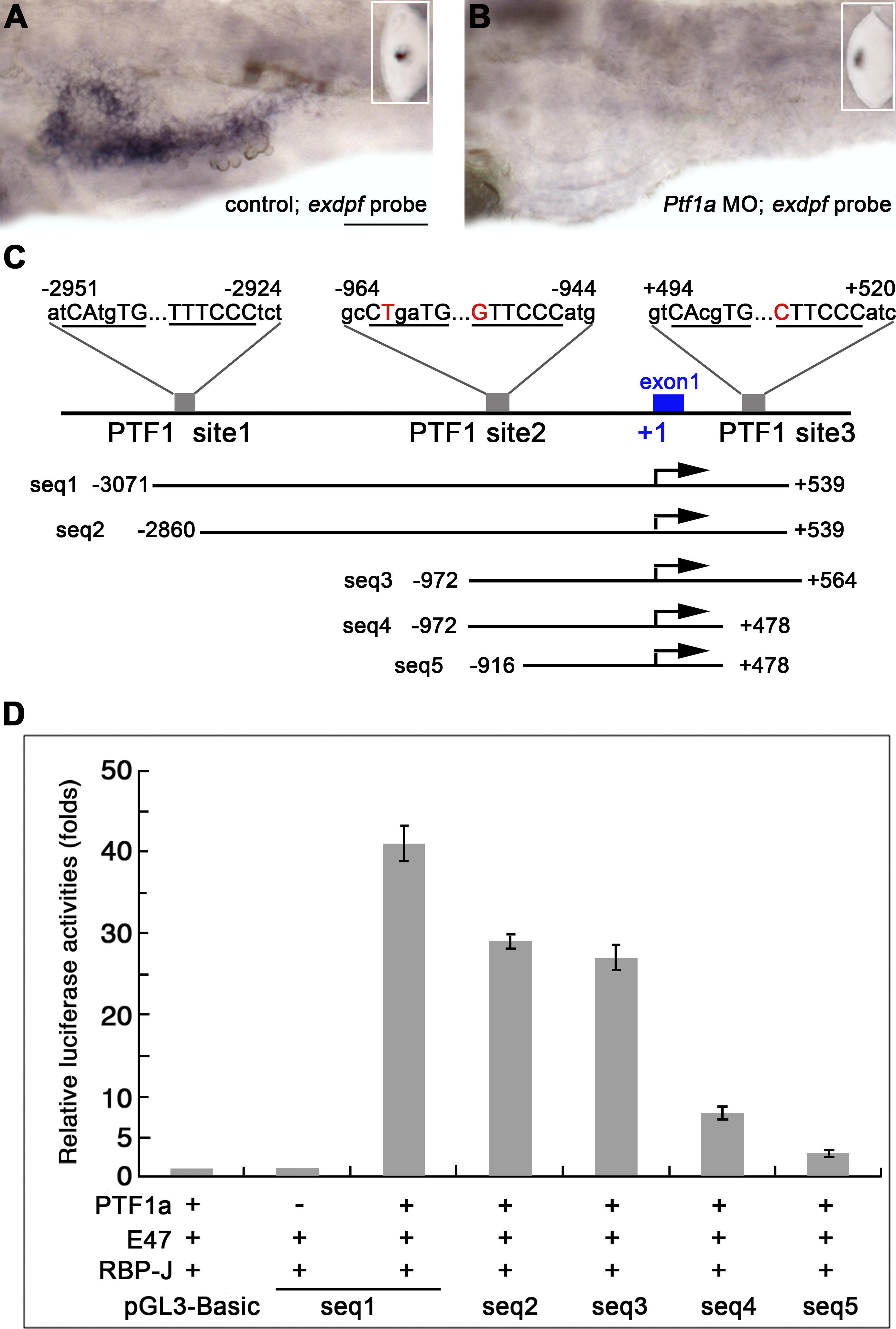
Fig. 4 Exdpf Expression Is Regulated by Ptf1a
(A and B) In situ hybridization using an exdpf probe in 3 dpf embryos. (A) An embryo injected with standard morpholino control. Inset: epiphysis expression. (B) An embryo inject with Ptf1a morpholino. Inset: epiphysis expression. Note that no exocrine expression of exdpf was detected. All embryos are shown in lateral view, anterior to the left. Scale bar: 50 μm.
(C) Locations and sequences of the three potential PTF1 binding sites in zebrafish exdpf gene and different exdpf promoter segments used in luciferase assay. The E-box and TC-box are underlined and capital letters indicate conserved nucleotides. The nucleotides that are not confirmed to the conserved nucleotides are in red. Dots indicate the spaces between the E-box and TC-box.
(D) Comparison of the activities of the segments containing different Ptf1 binding sites when transfected into 293 cells. These segments were inserted just before the ORF of luciferase gene. Luciferase reporter activity was adjusted for transfection efficiency and expressed relative to the promoterless pGL3-Basic vector. Error bars represent standard deviations.