
Fig. 3
- ID
- ZDB-IMAGE-090106-14
- Genes
- Publication
- Jiang et al., 2008 - Exdpf is a key regulator of exocrine pancreas development controlled by retinoic acid and ptf1a in zebrafish
- All Figures
- Figures for Jiang et al., 2008
|
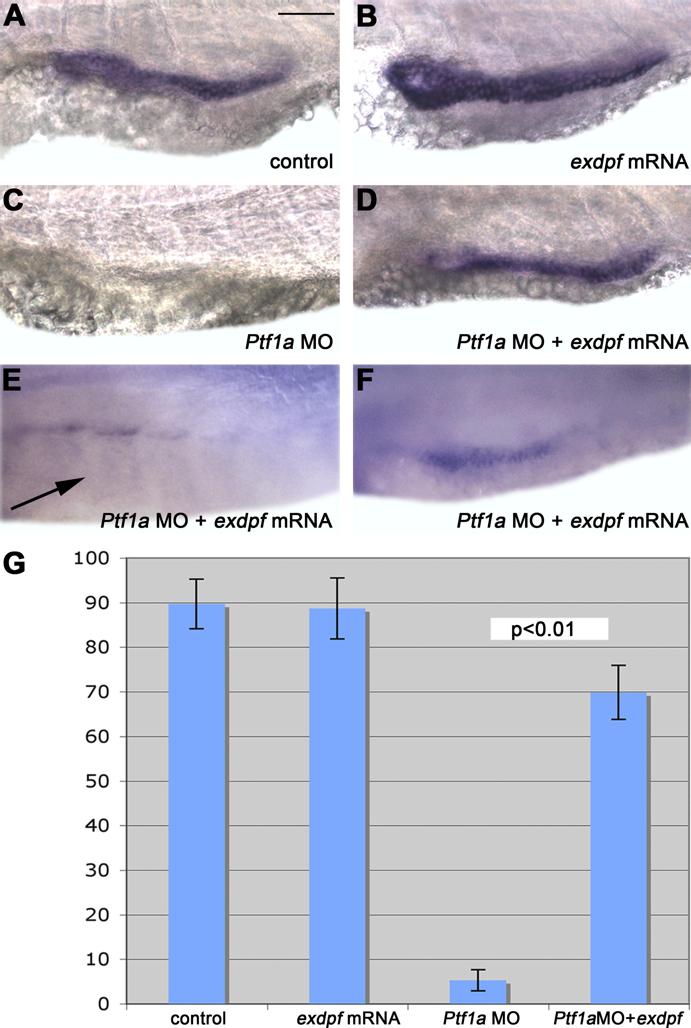
Fig. 3 Exdpf Restores Expression of the Exocrine Cell Marker in Ptf1a Morphants
(A–C) In situ hybridization using a carboxypeptidase A (cpa) probe in 3 dpf embryos. (A) A control embryo injected with water. (B) An embryo injected with 100 pg of exdpf mRNA. Note enhanced expression of cpa. (C) An embryo injected with 2 ng of Ptf1a morpholino. No expression of cpa was detected.
(D–F) Embryos injected with 100 pg of exdpf mRNA followed by 2 ng of Ptf1a morpholino. (D) Almost full restoration of cpa expression. (E) No detectable cpa expression in the presumed exocrine area (arrow). (F) Partial restoration of cpa expression. All embryos are shown in lateral view, anterior to the left. Scale bar: 50 μm.
(G) Percentage of embryos with cpa expression at 3 dpf. Y-axis represents mean ± SD.