
|
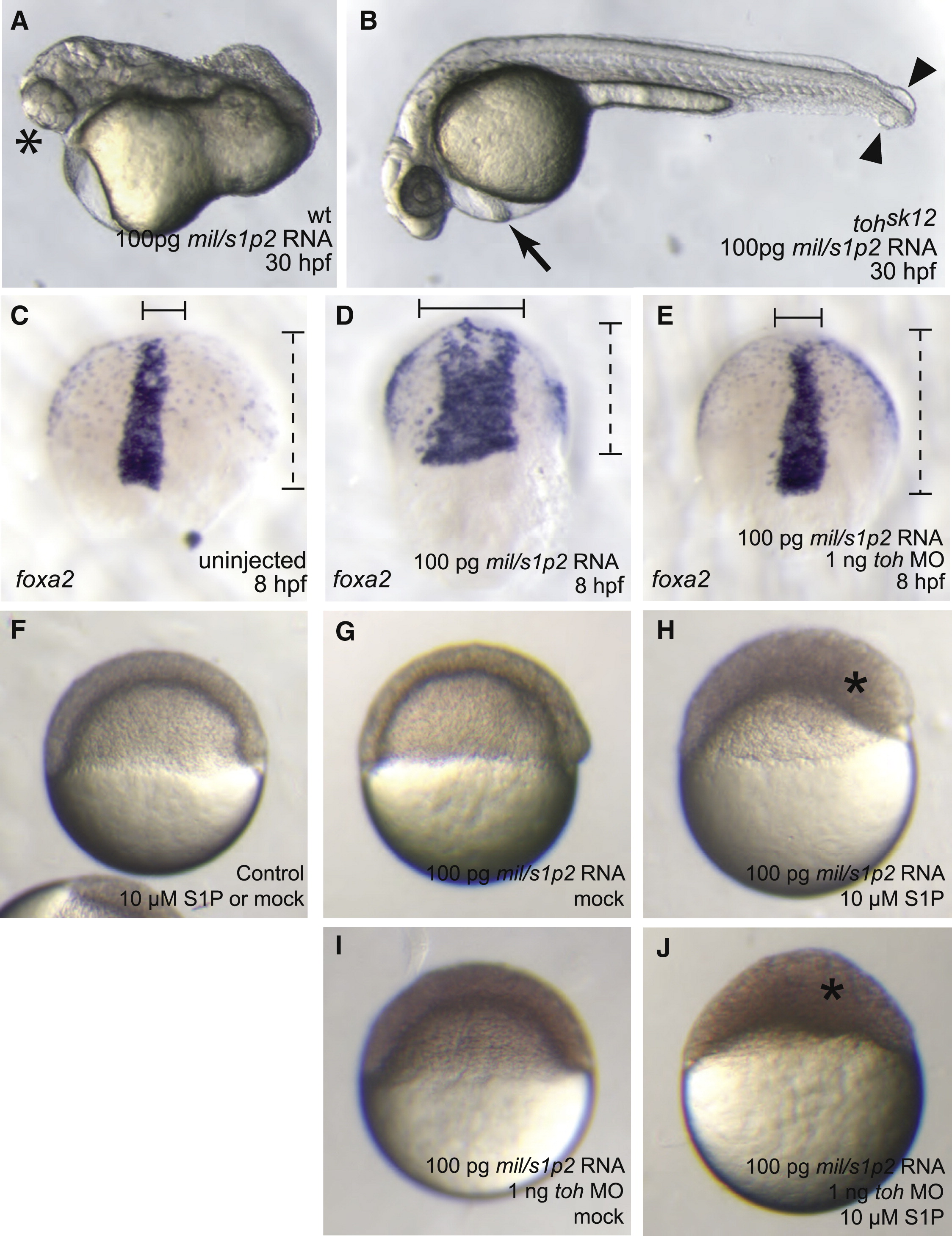
Fig. 4 Exogenous S1P Substitutes for Toh Function in Mil/S1P2-Overexpressing Embryos
(A and B) Lateral views with anterior to the left, of embryos overexpressing mil/s1p2 at 30 hpf. WT embryos injected with 100 pg of mil/s1p2 RNA (A) display a shortened body axis and cyclopia (asterisk). MZtohs8 mutants injected with 100 pg of mil/s1p2 RNA have no body-axis defects (B) but develop pericardial edema (arrow) and tail blisters (arrowheads).
(C–E) Visualization of axial mesoderm and endoderm by expression of foxa2 at late gastrula stages, dorsal views with anterior upwards. WT embryos overexpressing mil/s1p2 (D) have broadened axial mesoderm (solid line) compared to uninjected embryos (C). Progression of epiboly (dashed line) is also impaired in embryos overexpressing mil/s1p2 as compared to uninjected siblings. toh MO-injected embryos overexpressing mil/s1p2 (E) are indistinguishable from uninjected siblings (C).
(F–J) Lateral views of 5.5 hpf embryos injected with mock carrier solution or 10 μM S1P. Control embryos show no response to exogenous S1P (F). Embryos overexpressing Mil/S1P2 do not show more severe phenotypes when injected with mock carrier solution at 5 hpf (G). Mil/S1P2-overexpressing embryos injected with a 10 μM S1P solution at 5 hpf exhibit severe phenotypes (H), including an exaggerated thickened layer of cells at the animal pole (asterisk). Mil/S1P2-overexpressing embryos coinjected with toh MO show no phenotypes when injected with mock solution (I) and resemble controls (F). When injected with a 10 μM S1P solution, Mil/S1P2-overexpressing embryos lacking Toh function show severe defects (J), including a thickened layer of cells at the animal pole (asterisk).