
|
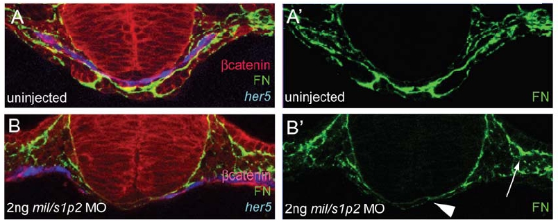
Fig. S4 Fibronectin Expression Is Affected in mil MO-Injected Embryos Confocal micrographs of cross sections of uninjected (A and A′) and injected with 2 ng of mil MO (B and B′) Tg(-0.7her5:EGFP)ne2067 embryos (GFP false colored blue) stained for β-catenin (red) to show cellular outlines and Fibronectin (green) at 20 hpf. Control embryos (A and A′) have endodermal cells (blue) spanning the midline (blue) and show strong Fibronectin deposition (green) under the endoderm and around the migrating precardiac mesoderm. mil MO injected embryos (B and B′) show gaps in the endodermal sheet (blue) and have considerably less Fibronectin deposition (green) at the midline (arrowhead), while lateral Fibronectin deposition (arrow) appears relatively unaffected.