
|
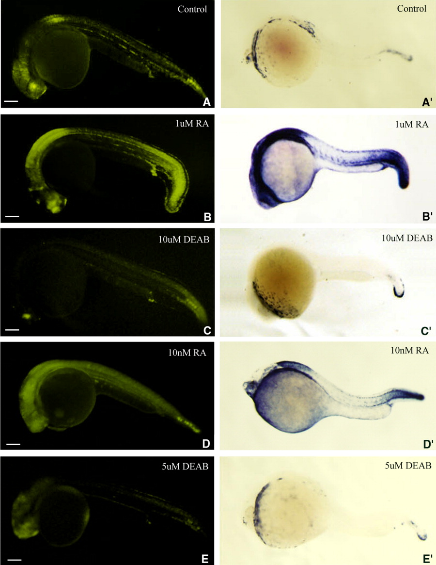
Fig. 6 Retinoic acid (RA) inducibility of enhanced yellow fluorescent protein (eYFP) expression in transgenic zebrafish embryos is similar to that of cyp26a1. B-E′: The transgenic embryos produced by a female wild-type zebrafish mated with a male transgenic zebrafish were treated with 1 μM RA (B,B′) or 10 μM DEAB (C,C′) for 6 hr starting from 18 hours postfertilization (hpf), or 10 nM RA (D,D′), or 5 μM DEAB (E,E′) for 24 hr starting from 0 hpf to examine whether the RA-induced expression of the transgene (B-E) is similar to that of cyp26a1 (B′-E′; revealed by whole-mount in situ hybridization) during zebrafish early development. A,A′: Untreated transgenic embryos were used as controls. Fluorescent images were photographed by a DP70 digital camera with 10-sec exposure time. Embryos are laterally viewed and positioned anterior left. In the embryos that were treated with 1 μM RA for 6 hr (B,B′), the greatly up-regulated expression was mainly seen in forebrain, anterior spinal cord, proctodeum, and whole tail (A,B,A′,B′). In the embryos treated with 10 nM RA for 24 hr treatment (D,D′), the up-regulated expression was almost in whole body but mainly seen in retina, anterior spinal cord and caudal notochord (A,D,A′,D′). When the embryos are treated with 10 μM DEAB for 6 hr (C, C′) or 5 μM DEAB for 24 hr (E,E′), the expressions of the transgene and cyp26a1 both are down-regulated mainly in retina, anterior spinal cord, and caudal notochord compared with wild-type embryos (A,A′). Scale bar = 100 μm.