
|
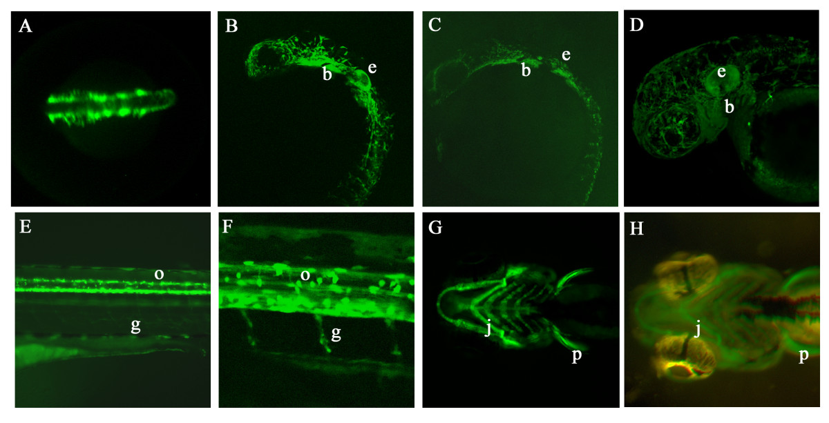
Fig. 3 GFP expression in germline transgenic sox10:GFP embryos. A. Dorsal view of head and anterior trunk of a Tg(-4725sox10:GFP)ba4 embryo at 16 hpf showing GFP expression in premigratory and early migrating neural crest cells. B, C. Lateral views of transgenic embryos from lines Tg(-4725sox10:GFP)ba4 (B) and Tg(-1252sox10:GFP)ba5 (C) to show similarities in GFP expression pattern at 24 hpf. (e ear, b branchial arches). D. Lateral view of head of a Tg(-4725sox10:GFP)ba4 embryo at 48 hpf showing extensive GFP expression throughout cranial pigment cells, ear (e) and branchial arches (b). E, F. Lateral views of the trunk of Tg(-4725sox10:GFP)ba4 embryos expressing GFP in oligodendrocytes (arrow) and Schwann cells (arrowhead). G, H. Ventral views comparing GFP expression in the jaw cartilage (j) and pectoral fins (p) of transgenic embryos from Tg(-4725sox10:GFP)ba4 (F) and Tg(-1252sox10:GFP)ba5 (G) at 96 hpf.