
|
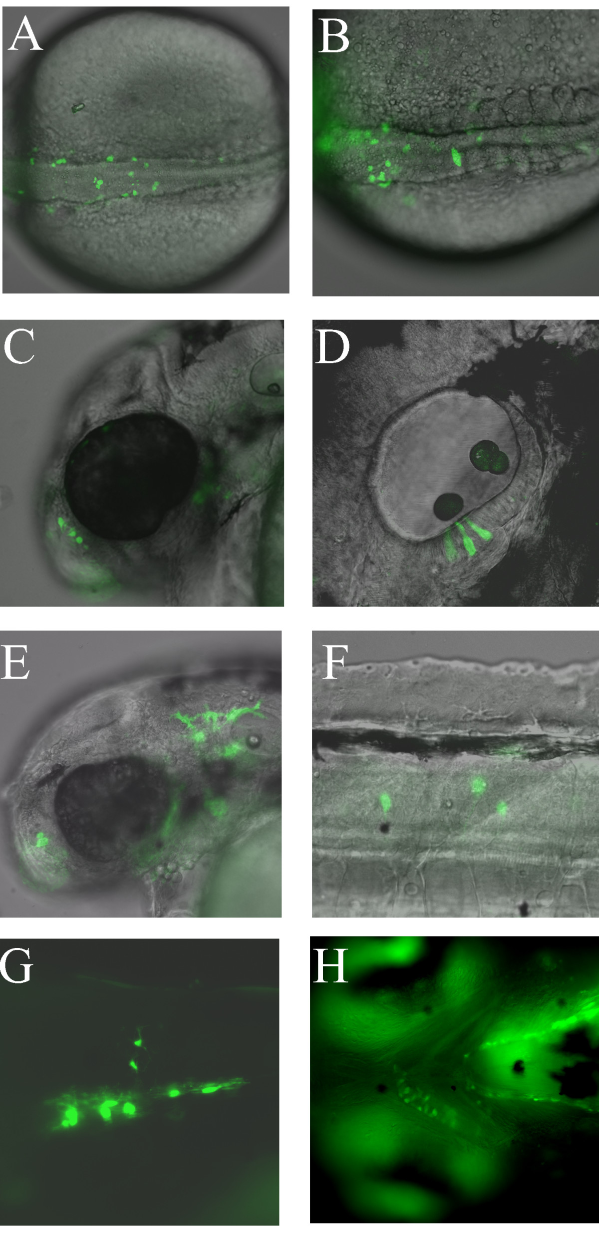
Fig. 2 Examples of mosaic GFP expression from transient transgenic sox10:GFP zebrafish injected with psox10-1252:GFP plasmid at the 1-cell stage. A, B. Dorsal views of two separate examples of pre-migratory neural crest GFP expression at the 20-somite stage. C. GFP expression in nasal cells at 48 hpf. D. GFP expression in otic epithelium at 48 hpf. E. GFP expression in xanthophores at 48 hpf. F. GFP expression in interneurons at 48 hpf. G. GFP expression in oligodendrocytes in trunk spinal cord at 96 hpf. H. Jaw cartilage expression at 96 hpf. All panels contain fluorescent images from live embryos (overlaid on DIC images in A-F). Orientation is with anterior to the left, dorsal uppermost except A, B (dorsal) and H (ventral).