
Image
|
Figure Caption
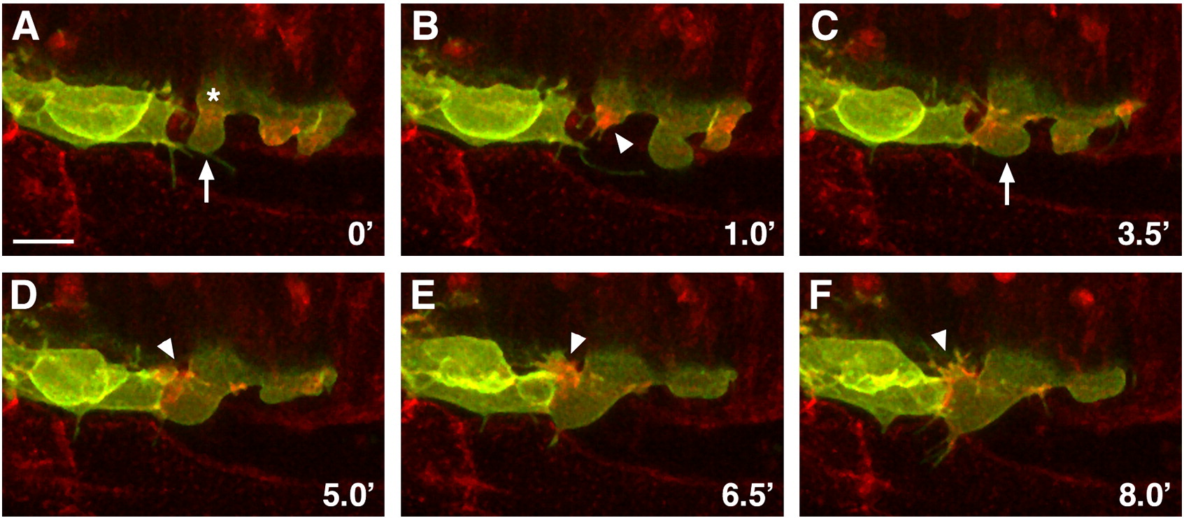
Fig. 5 Actin dynamics in blebs and lamellipodia in NCCs during EMT. Time-lapse sequence of NCCs at the edge of the neuroepithelium in embryos injected with sox10:CAAX-GFP DNA and RFP-UtrCh mRNA. (A) Cell marked with asterisk extends a bleb (arrow). (B) The bleb fills with actin signal upon retraction (arrowhead). (C) The cell blebs again (arrow). (D–F) The cell moves into the bleb while forming actin-filled lamellipodium and filopodia (arrowheads). Images are confocal projections. Dorsal views, anterior left. Scale bar = 10 μm.
Acknowledgments
This image is the copyrighted work of the attributed author or publisher, and
ZFIN has permission only to display this image to its users.
Additional permissions should be obtained from the applicable author or publisher of the image.
Reprinted from Developmental Biology, 324(2), Berndt, J.D., Clay, M.R., Langenberg, T., and Halloran, M.C., Rho-kinase and myosin II affect dynamic neural crest cell behaviors during epithelial to mesenchymal transition in vivo, 236-244, Copyright (2008) with permission from Elsevier. Full text @ Dev. Biol.