
|
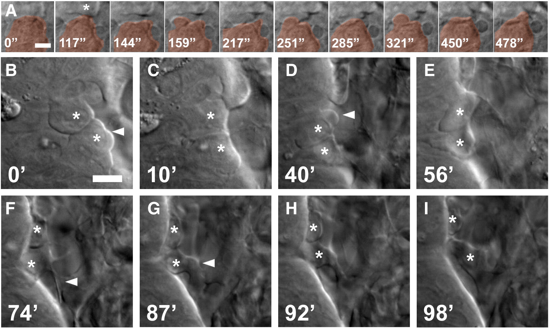
Fig. 3 Blebbing and filopodial protrusion occur during the NCC EMT. (A) Time-lapse sequence showing loss of cell adhesion and onset of blebbing in cell pseudocolored orange. Asterisk indicates a neighboring cell. Time is in seconds. (B–I) Time-lapse sequence showing dorsal view of NCCs undergoing EMT at the edge of the neuroepithelium. NCCs undergo blebbing (arrowheads in B, D) followed by migration out of the neuroepithelium accompanied by filopodial and lamellipodial extensions (arrowheads in F, G). Dorsal views of the basal edge of rhombomere 2/3, anterior is up, embryo is approximately 17 hpf. Time is in minutes.
Reprinted from Developmental Biology, 324(2), Berndt, J.D., Clay, M.R., Langenberg, T., and Halloran, M.C., Rho-kinase and myosin II affect dynamic neural crest cell behaviors during epithelial to mesenchymal transition in vivo, 236-244, Copyright (2008) with permission from Elsevier. Full text @ Dev. Biol.