
|
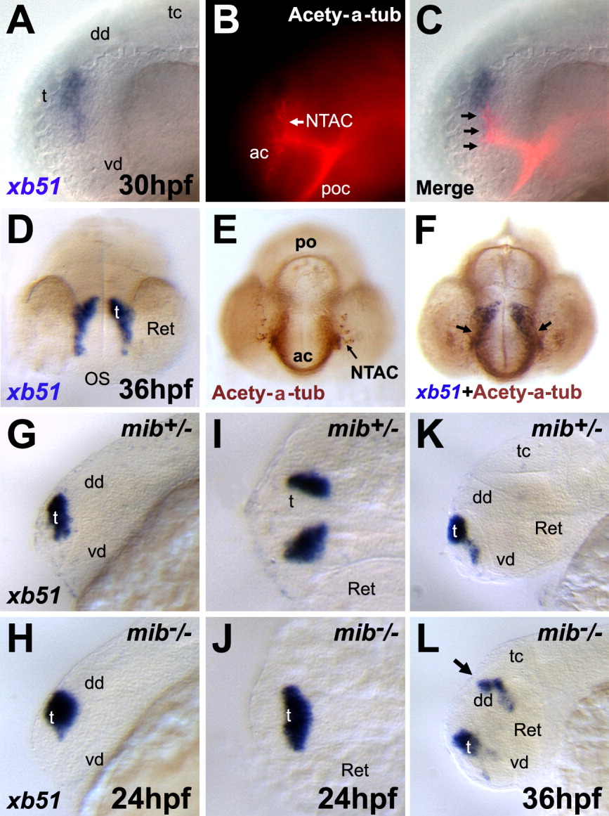
Fig. 4 Expression of xb51 in neuronal cells in the forebrain. Lateral (A-C, G, H, K, L), frontal (D-F), and dorsal views (I, J). Anterior to the left (A-C, G, H, K, L). A, D: Embryo hybridized with an xb51 riboprobe at 30 hpf (A) and 36hpf (D). B, E: Embryo stained with an anti-acetylated α-tubulin antibody, showing primary axon tracts. C, F: Double staining of embryos with xb51 and the anti-acetylated α-tubulin antibody. The xb51-expressing domain is very close to the region from where the anterior commissural axons (ac) originate (arrows). G-L: Expression of xb51 in a neurogenic mutant, mind bomb. The bilateral expression domains of xb51 in wild-type embryos (I) are medially fused in mind bomb mutant embryos at 24 hpf (J). At 36 hpf, xb51-expressing cells are ectopically detected in the midbrain of mind bomb mutant embryos (arrow). ac, anterior commissure; mlf, medial longitudinal fasciculus; NTAC, nucleus of the tract of the AC; po, posterior commissure; poc, postoptic commissure; sot, supraoptic tract; tpoc, tract of the postoptic commissure.