
|
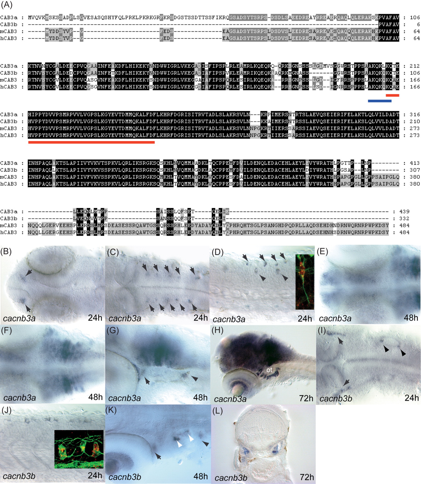
Fig. 3 Zebrafish cacnb3a and cacnb3b genes. A: Alignment of proteins encoded by zebrafish and mammalian CACNB3 genes shows that they are highly homologous to each other. The red bar underlines the β interaction domain (BID). The blue bar underlines the AKQKQKQ/S/V motif that is conserved in β1, β3, and β4. B: Dorsal view of the head showing that cacnb3a is expressed in two groups of cells (arrows) in the forebrain adjacent to the olfactory placodes at 24 hpf. C: Dorsal view of the hindbrain showing the expression of cacnb3a in discrete bilateral groups of cells (arrows) that may represent reticulospinal neurons at 24 hpf. D: Lateral view of the spinal cord showing cacnb3a is expressed in cells in the dorsal cord likely to be Rohon-Beard neurons (arrows) and commissural neurons (arrowheads) at 24 hpf. Inset from of an embryo labeled with the cacnb3a riboprobe (red) and anti-acetylated-α-tubulin (green) showing a cacnb3a-expressing commissural neuron extending a ventrally-directed axon. E: Dorsal perspective of the head showing cacnb3a is expressed diffusely in the brain at 48 hpf. F: Dorsal view of the hindbrain showing that at 48 hpf cacnb3a is strongly expressed by discrete groups of cells in rhombomeres 4-6 based upon the location of the otocyst. G: Lateral view of the hindbrain showing expression of cacnb3a in the trigeminal ganglion (arrow) and posterior lateral line ganglion (arrowhead) as well as the cells in rhombomeres at 48 hpf. H: Lateral view of 72 hpf embryo showing strong expression of cacnb3a in the brain, the retina and the cells ventral to the otocyst (ot). I: Dorsal view of the hindbrain at 24 hpf showing cacnb3b expression in the trigeminal ganglion (arrows) and in two groups of cells likely to be sensory neurons in the otic vesicle (arrowheads). J: Lateral view of trunk at 24 hpf showing cacnb3b expression in dorsal cells likely to be Rohon-Beard neurons in the spinal cord. Inset from an embryo labeled with the riboprobe for cacnb3b (red) and anti-acetylated-α-tubulin (green) showing that the dorsal cells express cacnb3b and extend longitudinal axons in the dorsal spinal cord consistent with them being Rohon-Beard neurons. K: Dorsal view at 48 hpf showing cacnb3b expression in the retina, trigeminal ganglion (arrow), otic cells (white arrowhead) and posterior lateral line ganglion (black arrowhead). L: Transverse section of 96 hpf embryo showing cacnb3 expression within the otocysts. Anterior is left in (B-K).