
|
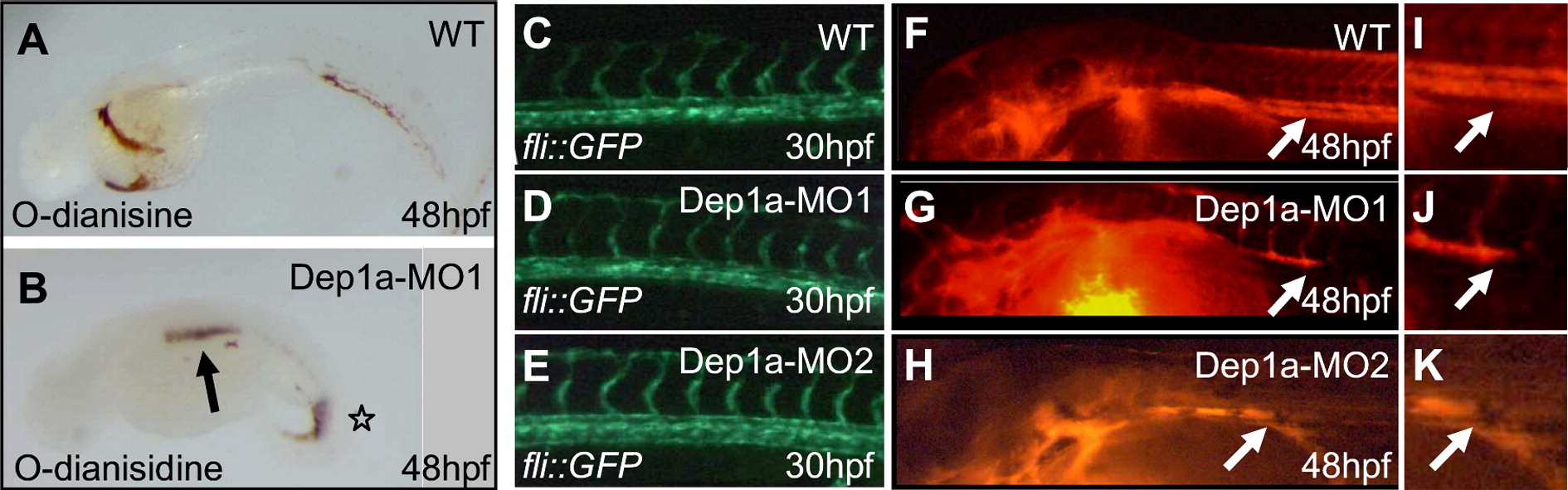
Fig. 3 Dep1a-MO induced blood circulation defects, but vasculogenesis appeared normal. O-dianisidine staining of hemoglobin was done on wild type (A) and Dep1a-MO1 (B) injected embryos at 48 hpf. The arrow indicates hemorrhages at the aortic bifurcation and the asterisk blood accumulation around the posterior cardinal vein. Fli1a::egfp1 transgenic zebrafish embryos were injected with Dep1a-MO1 (D) or Dep1a-MO2 (E) at the 1-cell stage and the vasculature was visualized at 30 hpf. A non-injected fli1a::gfp1 embryo served as a control (C). Microangiography was done by injection of rhodamine-conjugated dextran into the heart of embryos. Circulation of the dye was visualized using a fluorescence microscope. Non-injected control (F), Dep1a-MO1 injected (G) and Dep1a-MO2-injected embryos (H) all at 48 hpf are depicted here. (J–K) Magnifications of the area where circulation is blocked (arrow).
Reprinted from Developmental Biology, 324(1), Rodriguez, F., Vacaru, A., Overvoorde, J., and den Hertog, J., The receptor protein-tyrosine phosphatase, Dep1, acts in arterial/venous cell fate decisions in zebrafish development, 122-130, Copyright (2008) with permission from Elsevier. Full text @ Dev. Biol.