
|
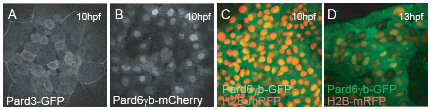
Fig. S1 Localization of Pard6γb prior to 24 hpf. (A–D) Images of live wildtype embryos. Embryos were injected with pard3-gfp, pard6γb-mch, pard6γb-gfp, and/or h2b-mrfp mRNA as indicated. (A) At 10 hpf, Pard3-GFP localizes primarily to the cytoplasm and cell membranes. (B) At 10 hpf, Pard6γb-mCh localizes to the nucleus, cytoplasm and cell membranes. [No nuclear localization signal was found in the Pard6γb-GFP fusion protein]. (C) At 10 hpf, Pard6γb-GFP co-localizes with H2B-mRFP in the nucleus. (D) At 13 hpf, Pard6γb-GFP begins to change its localization pattern. It localizes primarily to the cytoplasm and cell membranes.
Reprinted from Developmental Biology, 324(1), Munson, C., Huisken, J., Bit-Avragim, N., Kuo, T., Dong, P.D., Ober, E.A., Verkade, H., Abdelilah-Seyfried, S., and Stainier, D.Y., Regulation of neurocoel morphogenesis by Pard6gammab, 41-54, Copyright (2008) with permission from Elsevier. Full text @ Dev. Biol.