
|
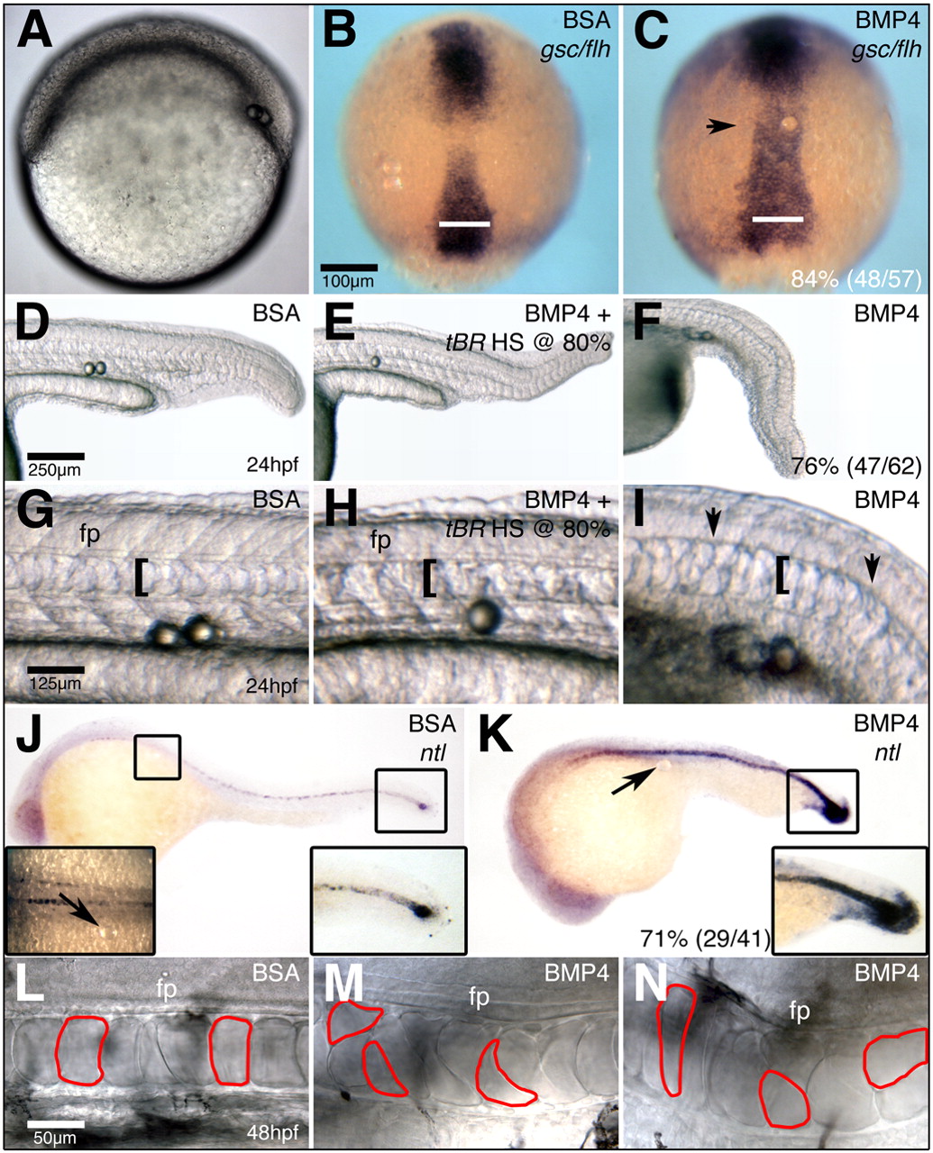
Fig. 7 A requirement for Bmp4 on the dorsal side of the gastrula. (A) Bead implantation on the dorsal side of the gastrula. (B,C) gsc and flh expression in 90% epiboly embryos that have had BSA- (B) or Bmp4- (C) soaked beads implanted into axial tissue. (D,F) The notochord of 24 hpf embryos implanted with BSA- (D) or Bmp4- (F) soaked beads. (E) tBR embryos implanted with Bmp4-soaked beads and heat-shocked at 80% epiboly. (G-I) High magnification images of the notochord in D-F. (J,K) ntl expression in 27 hpf embryos implanted with BSA- (J) or Bmp4- (K) soaked beads. (L-N) High-magnification images of the notochord at the level of the hind yolk extension of 48 hpf embryos that were implanted with BSA- (L) or Bmp4- (M,N) soaked beads. Floor plate is out of the focal plane in N. (B,C) Dorsal views, with anterior towards the top. Bars in B,C indicate the width of chordamesodermal domain at its widest point in the control embryo in B. All other views are lateral views, with anterior towards the left. Arrows in C,J,K indicate the position of the implanted bead. Brackets and arrows in G-I indicate the width of the notochord and absence of floorplate, respectively. Red outlines in L-N indicate notochord cell morphology.