
|
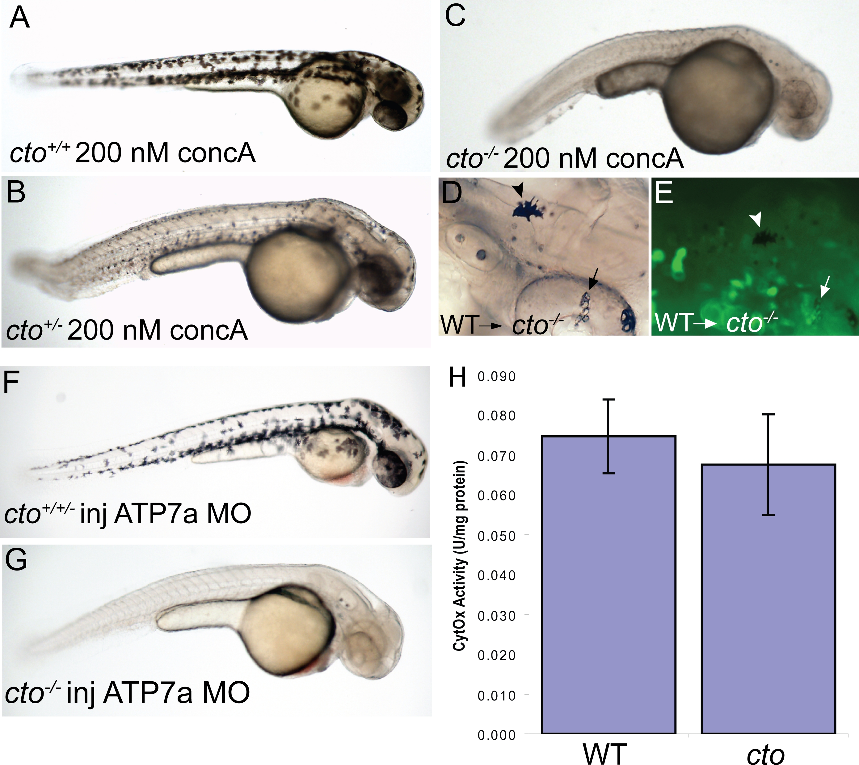
Fig. 5 cto contains a concanamycin A sensitive, cell autonomous defect which affects secretory pathway copper transport.
(A–C) cto gene dosage alters sensitivity to concanamycin A (concA) an inhibitor of Atp6. Wild-type fish have no phenotypic response when incubated in 200 nM concanamycin A beginning at 24 hpf (A). Embryos heterozygous for cto are sensitive to this same dose of concA, resulting in punctate melanocytes (B). ConcA exacerbates the phenotype of catastrophe homozygotes resulting in total loss of pigmentation and increased degenerative appearance (C). (D–E) The defect in cto is cell autonomous both in epidermal and retinal pigment epithelial cells. Wild-type, GFP-positive cells were transplanted into cto mutant embryos at the 1000 cell stage and allowed to develop to 48 hpf. Robustly pigmented melanocytes with normal size and shape can be seen sparsely distributed throughout the epidermis (D, arrowhead) and retinal pigment epithelium (arrow, D). The epidermal melanocyte does not have visible GFP but is not surrounded by GFP-positive cells (E, arrowhead). The RPE cells have a central area of GFP-positivity (E, arrow) Other areas are GFP positive without melanin pigment. (F–G) cto homozygotes but not heterozygotes or wild-type embryos are sensitive to atp7a morpholino injection. At a sensitizing dose of morpholino that does not affect wild-type/heterozygotes (F), homozygous cto embryos lose all pigmentation (G). (H) Cytochrome c oxidase activity is not reduced in cto embryos. Activity was normalized to protein levels in each sample. Three independent samples were prepared from three groups of embryos and the standard deviation of the three experiments is shown.