
Image
|
Figure Caption
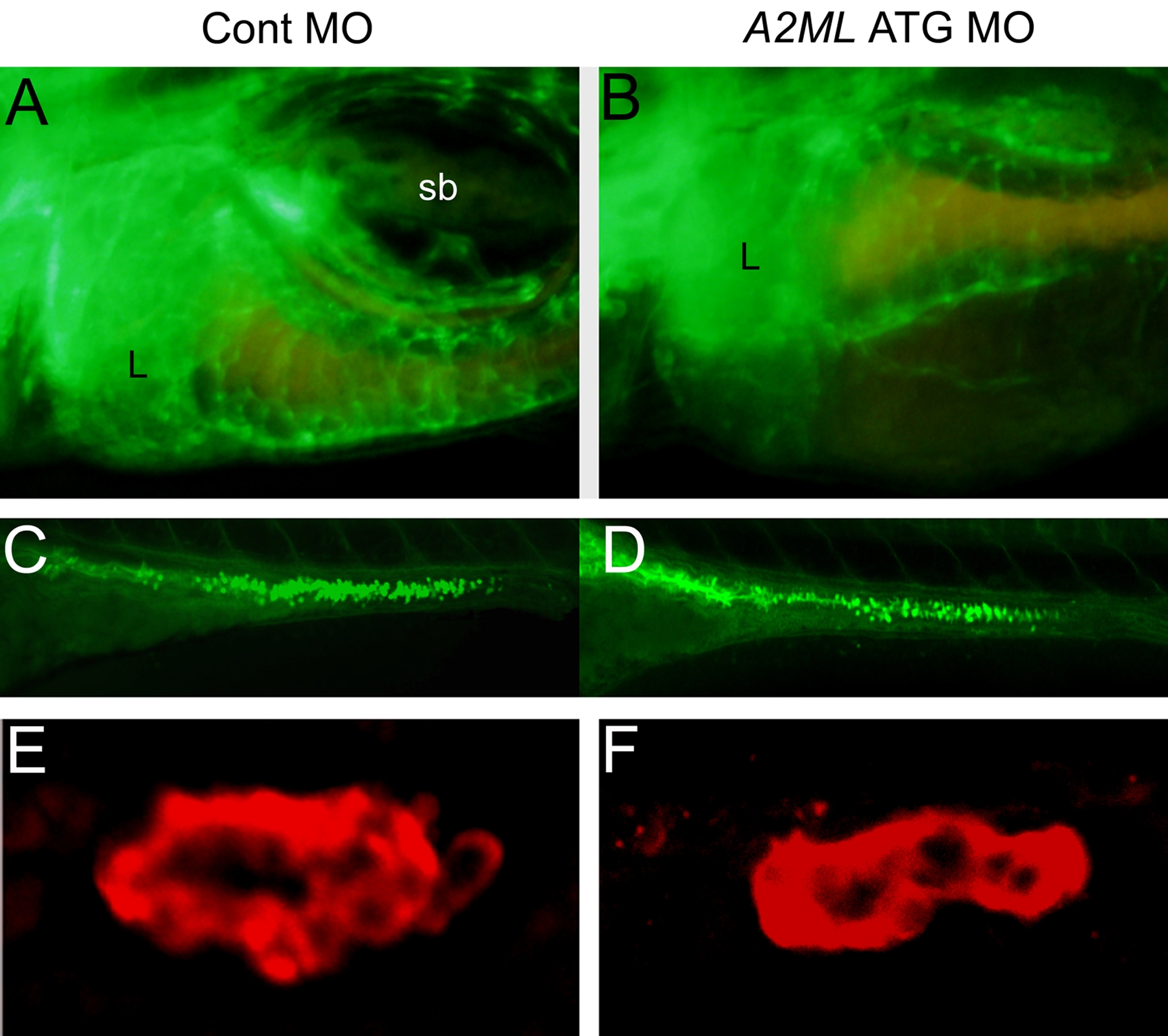
Fig. S2 Development of blood vessels, goblet cells, and exocrine pancreas. Embryo stages are 5 dpf for A–D, and 4dpf for E–F. A–B. Blood vessel formation of control MO (A) and A2ML MO (B) injected embryos as visualized in fli1-gfp Tg embryos. C–D. Confocal images of goblet cells in intestine were obtained using fluorescein-conjugated wheat germ agglutinin in control MO (C) and A2ML MO (D) injected embryos. E–F. Confocal images of immunoreactive carboxypeptidase A showing exocrine cells in the pancreas in cont MO (E) and A2ML MO (F) injected embryos. L, liver.
Figure Data
Acknowledgments
This image is the copyrighted work of the attributed author or publisher, and
ZFIN has permission only to display this image to its users.
Additional permissions should be obtained from the applicable author or publisher of the image.
Full text @ PLoS One