
Image
|
Figure Caption
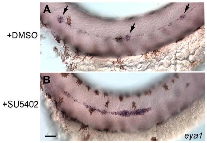
Fig. S2 Abrogation of Fgf Signaling Leads to a Primordium Migration Defect Very Similar to the Defect Observed in apcmcr Mutant Embryos
(A) In situ hybridization with eya1 at 38hpf. Primordium migration occurs normally and neuromasts are deposited (arrows) in wt embryos that have been incubated in DMSO between 20-38 hpf.
(B) In wt embryos that have been incubated in DMSO and SU5402 the primordium stalls and fails to migrate to the tip of the tail. Scale bar represents 40μM.
Acknowledgments
This image is the copyrighted work of the attributed author or publisher, and
ZFIN has permission only to display this image to its users.
Additional permissions should be obtained from the applicable author or publisher of the image.
Reprinted from Developmental Cell, 15(5), Aman, A., and Piotrowski, T., Wnt/beta-catenin and Fgf signaling control collective cell migration by restricting chemokine receptor expression, 749-761, Copyright (2008) with permission from Elsevier. Full text @ Dev. Cell