
|
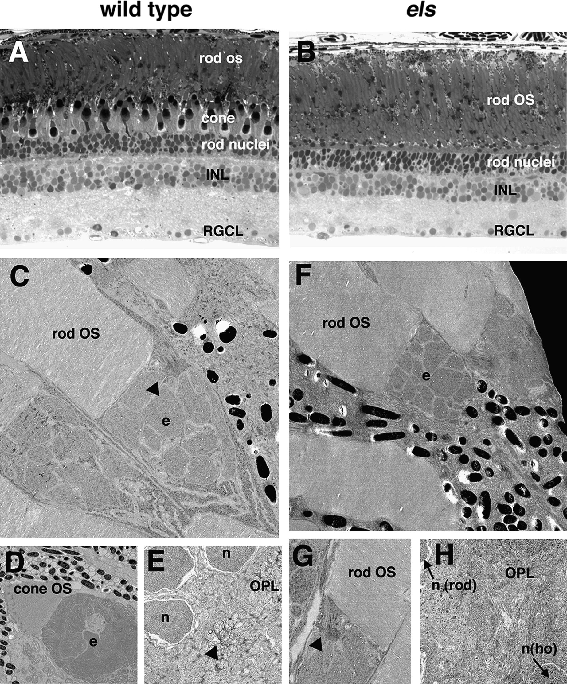
Fig. 5 Ultrastructures of photoreceptors in wild-type and els mutant at adult stage. (A–B) Semi-thin sections of wild-type (A) and the els mutant (B) retinas at 12 mpf. In the outer photoreceptor layer, the regions consisting of rod OS, cone cells and rod nuclei can be distinguished. In the els mutant retina, the region where cone cells are normally located appears to be replaced with the region consisting of the OS of rods, suggesting that cone cells are specifically eliminated in the els mutant. RGCL, RGC layer. (C) OS and ellipsoid (e) of rods in wild-type. Connecting cilia are observed (arrowhead). (D) OS and ellipsoid (e) of cones in wild-type. (E) OPL in wild-type. Dense-stained structures representing synaptic ribbons (arrowhead) are observed underneath rod nuclei (n) in the OPL. (F) OS and ellipsoid (e) of rods in the els mutant. These structures are normal. (G) Connecting cilia of rods in the els mutant. (H) OPL in the els mutant. Nuclei of rods (n (rod)) and horizontal cells (n (ho)) are shown at the upper left and bottom right corners of this image, respectively.
Reprinted from Mechanisms of Development, 125(11-12), Nishiwaki, Y., Komori, A., Sagara, H., Suzuki, E., Manabe, T., Hosoya, T., Nojima, Y., Wada, H., Tanaka, H., Okamoto, H., and Masai, I., Mutation of cGMP phosphodiesterase 6alpha'-subunit gene causes progressive degeneration of cone photoreceptors in zebrafish, 932-946, Copyright (2008) with permission from Elsevier. Full text @ Mech. Dev.