
|
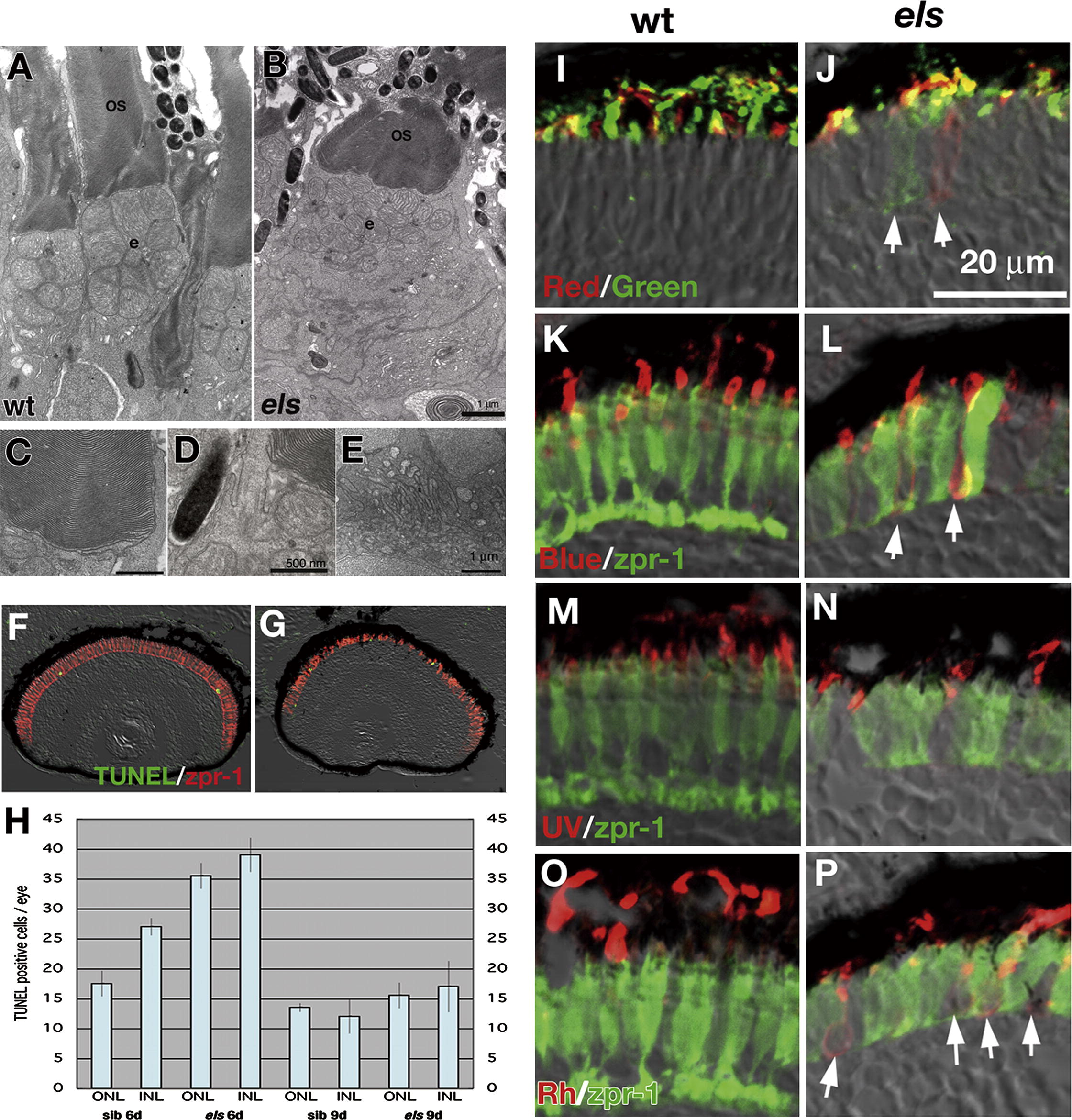
Fig. 3 The els mutant photoreceptors show abnormalities in cell shape, cell death and opsin localization in the OS. (A, B) EM analysis of wild-type (A) and els mutant (B) photoreceptors at 6 dpf. The OS develops from the apical surface of the photoreceptors and is composed of multiple-stacked membrane discs. Beneath the OS, mitochondria accumulate to form elipsoid (e). Although the global shapes of the OS and ellipsoid are deformed in the els mutant, their fine structure seems to be normal (B). (C–E) Ultrastructures of OS (C), connecting cilia (D) and synaptic region (E) in els mutant photoreceptor, which seem normal. (F, G) TUNEL (green) of wild-type and els mutant retinas at 6 dpf. The ONL are visualized by labeling with zpr-1 antibody (red), which stains the double-cone photoreceptors. (H) Histogram of number of dying cells per eye in ONL and INL of wild-type and els mutant larvae at 6 and 9 dpf. Light blue and error bars indicate average ± standard deviation. The cell death rates in the ONL and INL are higher in the els mutant than in the wild-type, and there is a significant difference between them at 6 dpf. (I, J) Labeling of wild-type (I) and els mutant (J) photoreceptor layers with antibodies against zebrafish red opsin (red) and green opsin (green). In some of the els mutant photoreceptors, both opsins fail to be localized to the OS but spread to the basal end of photoreceptors (arrows). (K, L) Labeling of wild-type (K) and els mutant (L) photoreceptor layers with anti-blue opsin antibody (red) and zpr-1 antibody (green). In the els mutant, blue opsin is mislocalized outside of the OS (arrows). (M, N) Labeling of wild-type (M) and els mutant (N) photoreceptor layers with anti-UV opsin antibody (red) and zpr-1 antibody (green). The mislocalization of UV opsin outside of the OS is not detected in the els mutant. (O, P) Labeling of wild-type (O) and els mutant (P) photoreceptor layers with anti-rhodopsin antibody (red) and zpr-1 antibody (green). In the els mutant, rhodopsin is mislocalized outside of the OS (arrows).
Reprinted from Mechanisms of Development, 125(11-12), Nishiwaki, Y., Komori, A., Sagara, H., Suzuki, E., Manabe, T., Hosoya, T., Nojima, Y., Wada, H., Tanaka, H., Okamoto, H., and Masai, I., Mutation of cGMP phosphodiesterase 6alpha'-subunit gene causes progressive degeneration of cone photoreceptors in zebrafish, 932-946, Copyright (2008) with permission from Elsevier. Full text @ Mech. Dev.