
|
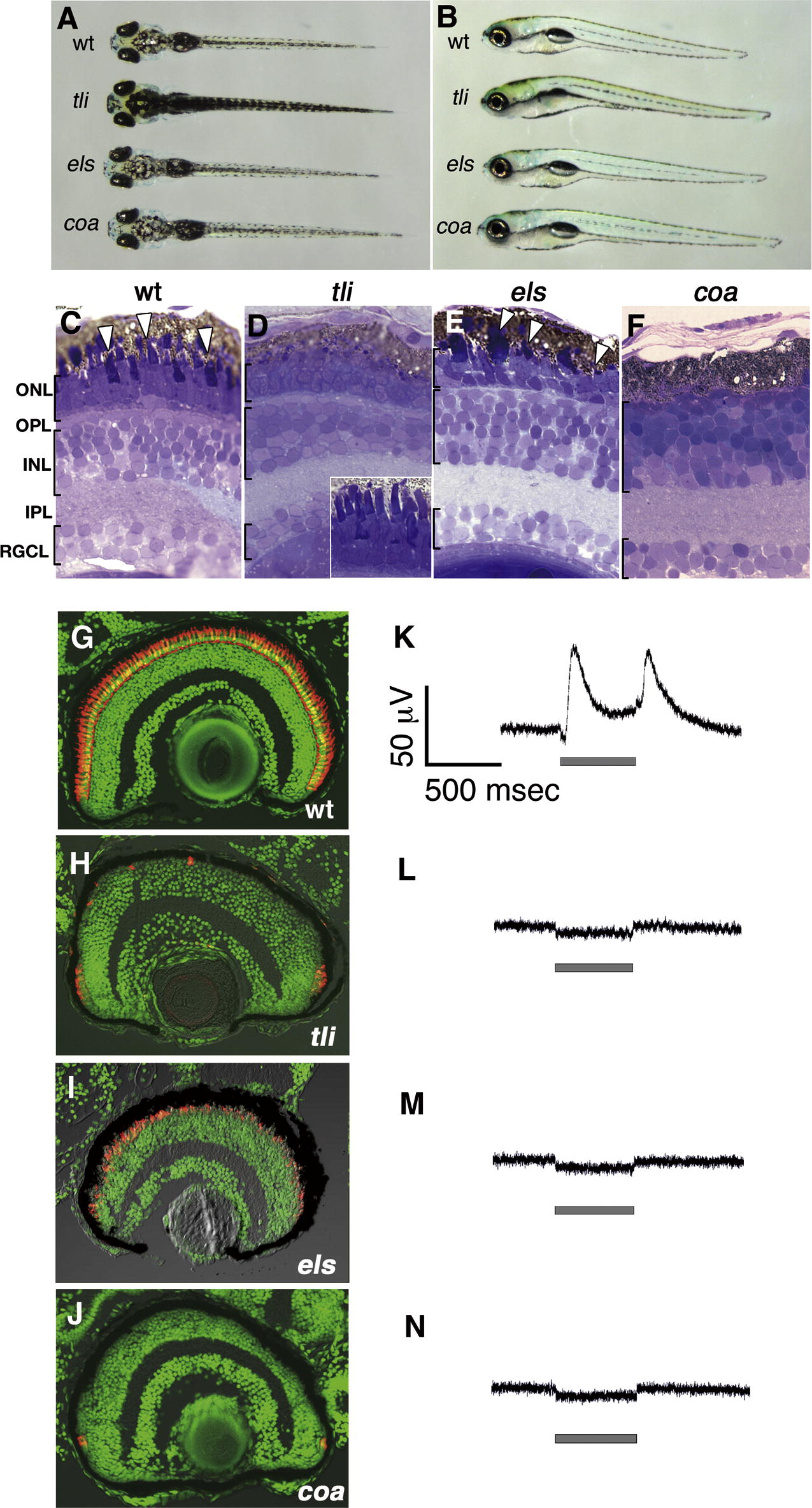
Fig. 1 Zebrafish OKR-defective mutants. (A, B) Dorsal (A) and lateral (B) views of wild-type, tli, els and coa mutant larvae at 7 dpf. The tli mutant larvae have expanded melanophores and no swim bladder. The els and coa mutant embryos have relatively normal body shape. (C–F) Plastic sections of wild-type (C), tli (D), els (E) and coa (F) mutant retinas. The stage examined was 7 dpf, except for the tli mutant that was examined at 5 dpf. In wild-type retina, three nuclear layers (RGC layer, INL, ONL) and two plexiform layers (IPL, OPL) are clearly evident (C). In the ONL, the photoreceptors differentiate and have a well-elongated OS, which is stained in dark blue (C, arrowheads). By contrast, in all three mutants, there are specific defects in the morphology of the photoreceptors. In the tli mutant, the nuclei of the ONL are observed to be normal, but the OS of the photoreceptors is specifically missing (D, the inset indicates wild-type 5 dpf photoreceptors). In the els mutant, photoreceptors are maintained, but the shape of the OS (arrowheads) is abnormal. In the coa mutant, both the ONL and OPL are absent. Furthermore, the horizontal cells are flat-shaped neurons normally located in the outer-most region of the INL, but are not observed in the coa mutant retinas. (G–J) Labeling of 8 dpf wild-type (G), tli (H), els (I) and coa (J) mutant retinas with zpr-1 antibody, which specifically labels zebrafish double-cone photoreceptors. All the nuclei are counterstained with Sytox-green (green). In wild-type retina, zpr-1-positive photoreceptors (G, red) form the ONL. In the tli mutant, zpr-1-positive photoreceptors are detected in the CMZ but almost absent in the central retina (H), suggesting that photoreceptors differentiate but degenerate in the tli mutant. In the els mutant, zpr-1-positive cells are maintained but their pattern is not regular (I). In the coa mutant, a few zpr-1-positive cells are observed in the region close to the CMZ, but they are completely absent in the central retina (J). (K–N) ERGs of 7 dpf wild-type (K), tli (L), els (M) and coa (N) mutant embryos in response to 500 ms light with an intensity of 50–60 μW/cm2. The wild-type embryo displays a typical response in ERG, which consists of a small negative a-wave and a large positive b-wave in ON-transient and a large positive d-wave in OFF-transient. However, all the three mutants show no response in ERG.
Reprinted from Mechanisms of Development, 125(11-12), Nishiwaki, Y., Komori, A., Sagara, H., Suzuki, E., Manabe, T., Hosoya, T., Nojima, Y., Wada, H., Tanaka, H., Okamoto, H., and Masai, I., Mutation of cGMP phosphodiesterase 6alpha'-subunit gene causes progressive degeneration of cone photoreceptors in zebrafish, 932-946, Copyright (2008) with permission from Elsevier. Full text @ Mech. Dev.