
|
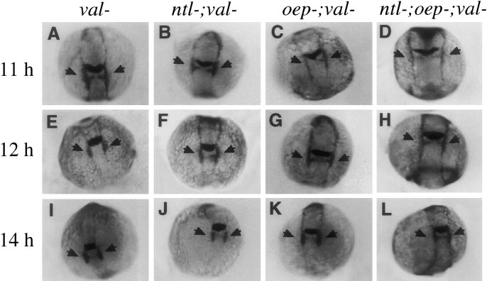
Fig. 7 Disruption of hindbrain development in val- embryos does not further delay expression of dlx-3. Shown are expression patterns of both dlx-3 and krox-20 at (A–D) 11 h, (E–H) 12 h, and (I–L) 14 h. Although embryos lacking val function cannot be morphologically distinguished at the early developmental stages examined here, val- embryos were identified unambiguously by virtue of their low levels of krox-20 expression in rhombomere 5. Expression of dlx-3 is normal in val- (A, E, I) and ntl-;val- embryos (B, F, J). Upregulation of dlx-3 in the otic regions (arrows) in oep-;val- (C, G, K) and ntl-;oep-;val- embryos (D, H, L) is delayed to roughly the same extent as in oep- and ntl-;oep- embryos with normal val function (see Fig. 3).
Reprinted from Developmental Biology, 206, Mendonsa, E.S. and Riley, B.B., Genetic analysis of tissue interactions required for otic placode induction in the zebrafish, 100-112, Copyright (1999) with permission from Elsevier. Full text @ Dev. Biol.