
|
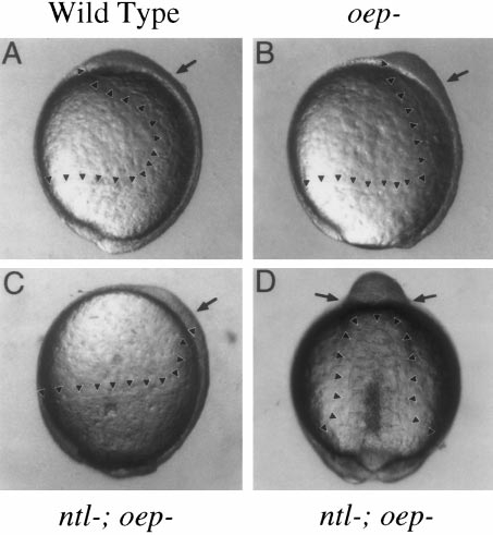
Fig. 3 Deficiency of mesendoderm in ntl-; oep- mutant embryos. Wild-type and mutant embryos were observed during gastrulation and neurulation to monitor movements of the hypoblast. Arrowheads mark the leading edges of the hypoblast, and arrows mark the approximate positions of the otic anlage along the anteroposterior axis. (A–C) Lateral views of embryos at 10.5 h, when otic induction normally begins. In wild-type embryos (A), axial hypoblast extends to just past the animal pole. Ventrolateral hypoblast has converged toward the axis, contributing to formation of paraxial mesendoderm. Epiboly is now complete and the tail bud is well formed. In oep- mutants (B), axial hypoblast extends only to the midbrain–hindbrain region, reflecting the deficiency in prechordal plate. Paraxial mesendoderm is clearly evident up to this region. Epiboly is about 95% complete. In ntl-;oep- mutants (C), axial hypoblast extends only to the anterior trunk region, and paraxial hypoblast is greatly reduced. At this time, there is essentially no hypoblast in the vicinity of the otic anlage. Epiboly is only 90% complete. From the lateral perspective, hypoblast movements appear normal in ntl- embryos (not shown). (D) Dorsal view of a ntl-;oep- double mutant at 13.5 h, around the time when otic induction begins in such embryos. The hypoblast forms a column of tissue that extends along the dorsal midline up to the anterior trunk region and gives rise to rudimentary somites. No hypoblast is evident anterior to the somites. Somites continue to mark the anterior limit of the mesoderm through at least 16 h, after which morphology becomes too disturbed to clearly resolve internal tissues (not shown). Necrosis of mesodermal tissues is variable, but often becomes evident during segmentation, as can be seen along the posterior midline of this specimen. The animal pole is oriented upward in all panels, and dorsal is to the right in A–C.
Reprinted from Developmental Biology, 206, Mendonsa, E.S. and Riley, B.B., Genetic analysis of tissue interactions required for otic placode induction in the zebrafish, 100-112, Copyright (1999) with permission from Elsevier. Full text @ Dev. Biol.