
|
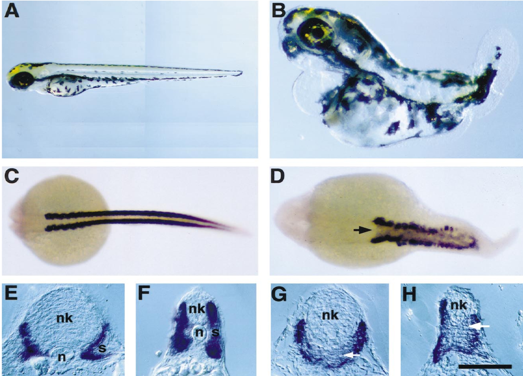
Fig. 5 Zebrafish embryos treated with 5-azacytidine lack tails and a differentiated notochord. (A) Wild-type embryos at 48 h of development have an elongated tail and possess a fully differentiated notochord in the trunk and tail. (B) Approximately 30% of embryos treated with 50 or 75 μM 5-azacytidine show reductions in the elongation of the tail, and loss or abnormal development of posterior somites. Structures anterior to the trunk appear to develop normally. (C, D) Transcripts of the muscle-specific α-tropomyosin gene are restricted to lateral bands along the body axis in 24-h wild-type embryos (dorsal view, C) but are also detected in the tissues that underlie the neural tube in 5-azaC-treated embryos (arrow, D). Transverse sections of (E, F) 24-h control and (G, H) 5-azaC-treated embryo hybridized with the α-tropomyosin probe. Control embryos show a large vacuolated notochord (n) directly beneath the neural keel (nk) and well-developed lateral somites (s), while sibling 5-azaC-treated embryos completely lack tissues resembling a notochord (arrow). (E and G, transverse sections posterior to hindbrain; F and H, transverse sections within posterior trunk/tail). Scale bar: A, 300 μm; B, 250 μm; C, 280 μm; D, 150 μm; E–H, 110 μm.
Reprinted from Developmental Biology, 206, Martin, C.C., Laforest, L., Akimenko, M.-A., and Ekker, M., A role for DNA methylation in gastrulation and somite patterning, 189-205, Copyright (1999) with permission from Elsevier. Full text @ Dev. Biol.